Volume 90%
Press shift question mark to access a list of keyboard shortcuts
Keyboard Shortcuts
Play/PauseSPACE
Increase Volume
Decrease Volume
Seek Forward
Seek Backward
Captions On/Offc
Fullscreen/Exit Fullscreenf
Mute/Unmutem
Seek %0-9
00:00
00:00
00:00
 

Chapters

Transcript


SPEAKER: Good morning, ladies and gentlemen. So it's always my great honor to be here again. I remembered the first time I attended this course in 2013. And this is my sixth time to be here. So last year I talked about full thickness resectioning. Two years ago, I talked about this, too. So this year what I will talk-- so today I will talk about from full thickness resectioning to true NOTES targeting extraluminal lesions.

So every year I perform the POEM case with Dr. Harrow, Dr. Towers [INAUDIBLE] in different three rooms. So for this course I think I will perform another-- maybe a difficult POEM case. As we know, for the development of the endoscope, there are about 200 years. From the reach the endoscope [INAUDIBLE] top flexible or fibro endoscope. But now we all use is a [INAUDIBLE] flexible scope. In 1960s, we began our endoscopy work in our center with a semi flexible endoscope in Shanghai.

So looking at this case, this patient once had an ERCP. After the ERCP, the patient had a severe necrotic pancreatitis. One month later, the patient had the high fever and the necrotic tissue in the upper-- in the [INAUDIBLE] peritoneal. We worked with the patient in the open surgery and we removed 1000 and 20 grams necrotic tissue-- and acute [INAUDIBLE] some terrible drainage in-- across the upper abdominal wall.

So three months later, the patient still had a fever, so from the abdominal sinus we put as a flexible scope-- catheter scope towards the patient's [INAUDIBLE] peritoneal and then removed the necrotic tissue with [INAUDIBLE] with [INAUDIBLE] and [INAUDIBLE] So that such a necrotic tissue was removed, and then that patient healed three months later. So the patient did not need the additional open surgery. So I think nowadays endoscope is not only the [INAUDIBLE] tool but also another surgical knife. We can use this knife to keep-- to perform some surgical procedures.

Based on endoscopic resectiioning from superficial disease when using the EMR technique, ESD technique to remove the precarious disease, to remove early cancers. From the submucosal layer-- because it emerges from the submucosal area, from the muscle area, we can even use the ESD and ESE or STER to remove the whole lesions. Even for some disorders in the stomach, in the esophagus, we can perform the POEM for the achalasia. So for the endoscopic minimally invasive resection we can now remove the lesions from the inner, from outer to the inner, some from the superficial to the deeper area.

This here-- this is a large [INAUDIBLE] hepatic flex [INAUDIBLE] So when you inject the saline into the supra-mucosal layer, you can see the lesion can be lifted very well. In the past, we always used the ESD technique to remove the whole lesion, just make an incision, and dissect the whole lesion from the submucosal layer. So nowadays I always choose a [INAUDIBLE] and 3 centimeters there to remove the whole lesion, and this is an EMR technique.

For larger lesions-- and for some large lesions, larger than 12 centimeters-- in the past we used a piecemeal EMR to remove the whole lesion. And this lesion is located in the duodenum-- from [INAUDIBLE] to the descending duodenum. So from other side to the anterior side, we removed the lesion piece by piece with [INAUDIBLE] So-- this-- now this lesion was removed-- was removed by more than 20 pieces.

So one piece is about one square centimeters, so the lesion was about larger than 10 centimeters. So when we do the piecemeal EMR we had to pick an advantage-- such that we could remove the whole lesion 5 centimeters, 10 centimeters, or 15 centimeters. But it is very difficult to give this loose specimens access or definite pathological diagnoses. Even, you can see, there are some tissue in [INAUDIBLE]. So the recurrence rate may be as high as 10% to 30%.

So on these circumstances, ESD technique was developed about 20 years ago in Japan. So this is recurrent lesion is in the middle part of the esophagus. So after the piecemeal EMR, when we checked the patient, in the middle of the part of the esophagus, we find-- we can find that this is-- a stricture ring. And this is a [INAUDIBLE] so the scope cannot pass through the stricture ring.

So first, we used the hook knife to make a longitudinal incision to relieve the-- to relieve the stricture ring, then the scope can pass through the stricture ring. And with the [INAUDIBLE] with the [INAUDIBLE] spread we can-- we can determine the [INAUDIBLE] recurrent or [INAUDIBLE] lesion. Then with ES technique, we can remove the whole lesions. So after the ESD technique, followed by the recurring lesion, this is a [INAUDIBLE]. So this part is a ESD [INAUDIBLE] and this part is the incision part.

So, for us nowadays, well, wor-- every day's work is becoming even difficulter. As you know, we have many, many of patients from all over China. So we can remove the superficial lesions with EMR, EPMR, or even ESD technique. How about submucosal tumors? This patient was five months old. During his mother's pregnancy, the patient was found to have a tumor in her stomach.

So what's this [INAUDIBLE] so was-- was he-- we'll say that the lesion was originated from the submucosal layer, no alcoholic. Is it a cyst? So with ESD technique, the whole lesion was removed from the patient's stomach. So at the end, the patient is very, very young, so the tissue was very, very [INAUDIBLE]. So the procedure time was only 10 minutes. So the last [INAUDIBLE] for this submucosal tumor was duplicate a stomach.

So nowadays, for tumors, even from muscle layer, we can use this technique, similar to the ESD [INAUDIBLE] endoscopic submucosal excavation for these deep lesions. So this-- this lesion was from the-- muscle layer is the rectum, just between the rectum and the vagina. So with a hook knife or with [INAUDIBLE] knife, such lesions can be removed with the muscle layer. So during the submucosal excavation for those-- from tumors from muscle layers, sometimes we can find the lesion was close to the serosal area, so on this circumstance, can we remove such lesions on endoscope?

So a new technique named full thickness resectioning was conducted in our center. So this is a residual lesion in the stomach. The patient once had a tumor resection in another hospital. During the procedure, they had massive bleeding, so the procedure was stopped. Several months later, the patient was referred to our hospital, like he was a patient, a ESD examination.

As you can see, the lesion was from the muscle layer. Just because that was a previous endoscopic resection, there always was a severe process in the submucosal layer. On this [INAUDIBLE] I just make a-- make the actual hole around the lesion, and then removed the whole lesions with the serosal layer. And this is the specimen after this-- after resection, this is the serosal layer. You can see the clear vessels in the serosal layer. So this is a true full thickness resection.

This In another case of the full thickness resection. This is of a loose tumor in the stomach. CT scan issued [INAUDIBLE] was actually extraluminal. So we just make a hole in this area, then put a scope outside of the stomach, and removed the whole lesion. And this is a GIST-- this is a GIST in the extraluminal growth.

So last year, we were performing another interesting case. So this is a [INAUDIBLE] tumor at the orifice of the appendix. ES showed the lesion was from the muscle layer. Can we remove such a lesion on colonoscopy? So, at the end, the lesion was located at the orifice of the appendix. So I decided to remove the whole lesion, including the appendix. So we make a full thickness resection around the [INAUDIBLE] tumor-- that is around the orifice of the appendix.

So during dissection, when we dissected the appendix from the mesenterium, we find that this is the appendic artery, and so we used the [INAUDIBLE] to stop the bleeding from the artery. Then we removed the whole lesion with the-- with the appendix. And the disease is a submucosal tumor in the orifice of the appendix. And this is the appendix. So I think this is an endo-- this-- this procedure is an endoscopic appendectomy. So I think these are the true NOTES. For the NOTES, I think comparing with a laparoscopic procedure, notice-- notice has the same procedures with a traditional laparoscopy procedure, but the access was different.

So recently we finished the first-- the first endoscopic gastrojejunestomy without a help [INAUDIBLE]. With just the opening-- opening the stomach with a little knife and the balloon, then put in the scope into-- out-- outside the-- outside of the stomach. With the forcep, we get the jejunum into the lumen-- into the gastric lumen. Then with several clips to fix the jejunum.

Now we use clips to affix the jejunum to the stomach. And then we open the jejunum with a little knife. And the last thing we insert, for a cover, the stent between the jejunum and the stomach. I think this is the endoscopic gastrojejunestomy, without help, I was at US guidance.

So nowadays, for the NOTES, we're just opening-- we just make an active or direct access through the stomach to then put a scope outside of the lumen, just to perform surgical procedures. This is a submucosal tumor. It looks like from the muscle area, but the CT scan showed that the lesion was totally extraluminal.

And that the lesion was close to the-- and this, on the right, is a kidney. So we just make an access through the stomach, and then we put a scope into the abdominal cavity. And we found that the lesion was close to the retroperitoneum. And the lesion was actually comes from retroperitoneum. And last up, [INAUDIBLE] for this lesion was access with the spleen.

Look at this case. This also looks like a submucosal tumor. Where you were removed-- the way we cut into the gastric wall, we cannot find the lesion. Where is the lesion? Actually the lesion was comes from the spleen. And this is a splenic cyst.

So recently we performed several cases of true NOTES, for the extraluminal lesion. This is a lesion located between the stomach and the liver. And it is a tumor. The tumor was about 1.5 centimeters. We make an opening in the stomach, put it forward into the abdominal cavity, and we found the lesion was located between the liver and the stomach. With [INAUDIBLE] knife, hook knife, and [INAUDIBLE] knife, the lesion was totally removed. At last we used several clips and a loop to close the access in the stomach.

So with that development of the full thickness resection and the tunneling endoscopy, we think, can we make a tunnel to remove the lesions outside of the-- as a [INAUDIBLE] outside of the stomach? So we can make a short tunnel always about 5 centimeters in the supra-mucosal layer, then put in a scope along the supra-mucosal tumor-- cut it up, cut the mucosa, cut the serosal tissue about the tumor, then put in a scope from the supra-mucosal [INAUDIBLE] to the extraluminal. Then remove the whole lesion. The last step of this procedure is the same to the POEM or to the STER.

So this is a lesion. It is also located between the stomach and the liver. So the lesion was around each junction. So with just a big turnover, and a cut to the muscle about the tumor. So when we find the tumor, we just rem-- dissected the tumor along the capture So this area, this is a tumor.

So when we-- when the tumor was dissected, we can use the basket or use the [INAUDIBLE] to remove the whole lesions from the tunnel, even from the patient's mouth. So the last step [INAUDIBLE] I was isolating it, so once normal. One month later, when we followed-- when we checked into the patient, in this area, so the tumor disappeared. And there was no recurrence.

Another procedure for us for NOTES is a reservoir of the gallbladder. So for the gallbladder stone in the past, we think, just cholecystectomy is the traditional procedure for those patients. Now we just choose some special patients to do the patient's gallbladder preserving [INAUDIBLE]

So the first for this procedure, first, we make an access in the stomach. Then when we find the gallbladder, make a hole in the fundus of the gallbladder. And then put a scope into the core-- in the lumen of the gallbladder. Then we use several clips-- several baskets to remove whole stones from the gallbladder lumen. In the past, we always used several clips to close up entrance in the gallbladder.

So this short video showed us the whole procedures of this-- whole procedures of the gallbladder, preserving [INAUDIBLE] So we make a hole in the anteroom. Cut out the muscle-- cut to the muscle layer. And with a hook knife, then we change to use a [INAUDIBLE] knife to make a big entrance. With the help of the cup, the scope can be put into the abdominal cavity. And this-- this is the gallbladder.

We use the hook knife, or [INAUDIBLE] knife to make a-- make a hole or make an access in the fundus of the gallbladder. The gallbladder wall is very thick in the fundus. And then now the scope can be put into the lumen in the gallbladder. And then we can find the several stones in the lumen. With this new basket, such stones can be removed, or take it out from the patient's mouth.

These are removed stones. And this is the mucosa in the gallbladder. And next we use clips to close the entrance in the gallbladder. Five to six clips are always needed. So at last I will use a clip and enter lobe to close the entrance in the stomach. So nowadays we began to use an over-stitch to close the entrance of the gallbladder.

This is another interesting case. The patient had a submucosal tumor in the stomach from the muscle layer. So first step, we removed the submucosal layer from the stomach, then from the [INAUDIBLE] we put the scope into the abdominal cavity. The gallbladder was full of stones. When that gallbladder was opened several stones come from the lumen into the abdominal cavity, then we removed the [INAUDIBLE] from the gallbladder from the abdominal cavity. When we-- at last we calculated the number of the stones, there was about 229 stones. This is a map [SPEAKING CHINESE] stones-- 229. So [SPEAKING CHINESE] gallbladder.

For some, [INAUDIBLE] stones in the neck of the gallbladder is very, very difficult to remove it with just the use of forcep to make the stone shorter-- to make the stone loose, then such stones can be removed. So we even can find some stones within the gallbladder wall. So when the stones was removed from the lumen of the gallbladder, we found some black points in the mucosa. What is these? I just opened these black point, then some black stones comes out. So this stone is in the gallbladder wall.

So last year we finished 30 cases of such a procedure. And from this month, every week we have one to-- one to two cases for the gallbladder reserve [INAUDIBLE] So those are stones that are removed from the patient's gallbladder through the [INAUDIBLE] access

So for the translumenal endoscopy, there are two accesses. First is direct, we think, is the full thickness resection and the [INAUDIBLE] on NOTES. Another [INAUDIBLE] is direct, and that's just [INAUDIBLE] with the submucosal [INAUDIBLE]

So, ladies and gentleman, full thickness resection and NOTES makes the transluminal procedure for extralumenal lesion possible. The future of the flexible endoscopy is not only operating in a natural orifice but also within and outside the gastrointestinal wall. Thank you very much for your kind attention.

[APPLAUSE]

Video

From Targeting EFTR to True NOTES: Targeting Extraluminal Lesions

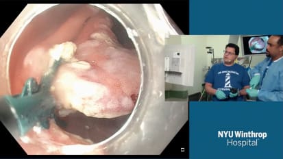
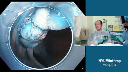
Ping-hong ZHOU, MD, FASGE, Zhongshan Hospital, China, provides a lecture on targeting extraluminal lesions. 

Related Presenters

Ping-Hong Zhou, MD.

Ping-Hong Zhou, MD

Doctor of MedicineGraduate SupervisorDeputy Director of Endoscopy CenterAssociate Professor of General SurgeryYouth Member of Chinese Society of Digestive EndoscopyMember of Endoscopy Branch of Chinese Medical Doctor AssociationMember ...

View full profile

ADVERTISEMENT

Related Videos