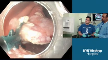
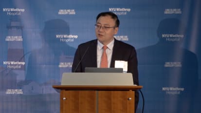
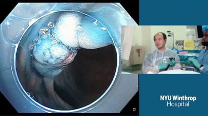
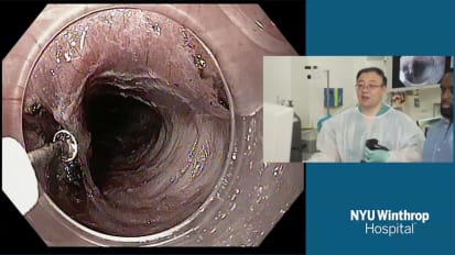
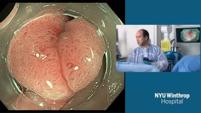
LUO XIAOBEI: Next, I'm going to share endoscopic full-thickness resection with mucosal preservation. Endoscopic treatment of gastric submucosa tumorous were usually done by ESD or EFTR. When perforation occurs, [INAUDIBLE] combined with endoclips, OTAC, or overstitch systems were usually applied to make the closure.
Today, I'm going to share a case in which the wound of EFTR was closed only by the normal mucosa over the SMT, combined with endoclips. We have developed a modified method called endoscopic mucosa-sparing lateral dissection technique for the removal of gastric SMT. First, markers were made with APC slightly exceeded the border of the tumor.
After submucosa injection, several small and short incisions were made on both sides of the borders. After that, the mucosa was cut over a semicircle, and the submucosal tissue was carefully dissected to separate the submucosal tumor with the gastric mucosa. When fully exposed, the submucosa tumor was carefully dissected.
If the tumor locate originates from that the deep layer of the muscularis propria, especially when it's a little extraluminal growth, full thickness resection was performed after reaching the serosa layer. When the tumor was fully resected, we placed back the intact mucosa, and used endoclips to clutch the small incisions together and drag the retracting mucosa. And then, finally, make the closure.
Next, I'm going to share a case of EFTR covered with mucosa to make the final closure. It's a 23-year-old female, and gastroscopy revealed a 15-millimeter submucosa tumor in the anterior of the greater curvature of the gastric antrum. EUS suggested that the tumor originated from the muscularis propria with a little extraluminal growth.
Here is the video of the EFTR with mucosal preservation. Markers were made with APC, and several short and small incisions were made on both sides of the markers. This helped to trace the origin position of the mucosa. And it also helps us to make the final closure easier and faster.
After the mucosa was cut over the semicircle, submucosa was dissected to separate the gastric mucosa and the tumor. Attention should be paid to avoid damaging the integrity of the mucosa. And then, when the tumor was fully exposed, the submucosa tumor was carefully dissected.
If that tumor originates from the deep layer of the muscularis propria, endoscopic full thickness resection was performed after reaching the serosa layer. So attention should be paid when doing the full thickness resection to not damage any of the abdominal organs.
So if you use the IT [INAUDIBLE] or the hook [INAUDIBLE], we will have to use some of the pullback technique. So when the tumor was fully resected, the intact mucosa was placed back to cover the surgical wound and the perforation. And the endoclips were used to clutch the small incisions together, drag the retracted mucosa, and make the closure.
To facilitate the closure, we can make full suction and then reduce gastric air to make the retained mucosa close to the wound. And use the tip off the transparent cap to unfold the retracted mucosa. Usually, we start to fire the first endoclip in the middle of the wood. And step by step, we use the intact mucosa combined with endoclips to completely close the wound and the perforation.
That's all of the procedure. And the operating time is about 60 minutes. En bloc resection was achieved without major post operative complications. And follow-up three months later revealed that the wound healed completely with several clips remaining.
In conclusion, endoscopic resection can be considered for small and large gastric submucosal tumors in selective cases. And the integrating of the mucosa should be emphasized. That wound of EFTR or perforation can be closed safely with the retained mucosa, combined with endoclips.
That's all for my presentation. Thank you for your attention and time.
SPEAKER 2: Thank you.
[APPLAUSE]