MALE SPEAKER: This video is discussing transoral robotic surgery of the base of tongue in a patient with a minor salivary-gland tumor. The teeth are protected with a Aquaplast Thermoplastic Protector to prevent injury of the teeth. It's very important in transoral robotic surgery to get the maximum exposure that you can of the tumor. And typically we do that with the help of the modified FK oral retractor.
This has value over a traditional laryngoscope in that we can usually get a wider view of not only the base of the tongue but also the surrounding landmarks for orientation. It's important to carefully understand the location of the tumor prior to placement of the laryngoscope, so studying the images is helpful.
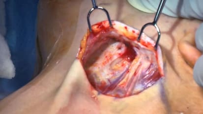
Feeling the tumor with the patient under the anesthesia prior to introducing the laryngoscope to gain a good mental impression of the boundaries of the tumor is important. With the oral tractor in place and suspended, ideally the majority of the tumor should be in view with some surrounding view of the epiglottis, lateral pharyngeal wall, and posterior pharyngeal wall.
On the side table it's helpful to have clipper pliers, other laryngoscope blades within access, and sections for the assistant. After placement of the oral retractor, the robot is docked.
Docking of the robot-- in this case, the SP version of da Vinci --consists of placement of the cannula. The endoscopic camera is placed first and then the robotic arms, consisting of graspers, bipolar cautery, unipolar cautery, needle drivers, as needed, are then placed.
The end of the cannula is placed approximately 7 to 8 centimeters from the incisors. This gives the SP instruments the ability to maximally expand before converging again at the working end, which should be at the level of the tumor. A monitor is in place for the assistant so that they have adequate view of the operative field.
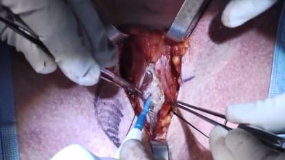
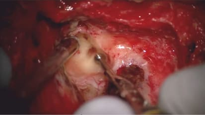
The surgical assistant plays a vital role in suctioning smoke and fluids, helping with retraction, placing clips for hemostasis, and even bipolar cautering. This view shows the right base of tongue tumor in view with the epiglottis and surrounding landmarks for orientation. The procedure is initiated with cuts through the mucosa, both laterally and medially, and usually anterior to the tumor to allow the tumor to drop further into the operative field.
These cuts are in place several centimeters from the obvious edge of the tumor, or the surgeon's impression of the edge of the tumor. The anterior cuts are critical. At this point we're going to skive beyond the interior edge of the tumor into the intrinsic tongue musculature. One of the pitfalls of this operation is making that cut too close to the tumor edge or even through the tumor edge, studying the preoperative radiographs and study of the tumor pre operatively can help prevent that.
The procedure proceeds with further cuts through the mucosa, further cuts through the intrinsic musculature, and assessment of vasculature that may need to be managed. The surgeon controls the camera with the help of the foot pedals and with the robotic instrument arms.
And both arms are available for manipulation of the tumor. A third arm can be placed for help with retraction in the tumor. The surgeon can switch back and forth between the second and third arm by depressing the foot pedals.
As the tumor drops further into view, it's important to continually assess the boundaries of the tumor. Cuts are made through the intrinsic tongue musculature, which is very obviously different from the tumor because it has contractile ability. So as these cuts are made, the muscle fibers will spread apart very easily. If the muscle fibers aren't spreading apart easily, the surgeon should be concerned that they're within the parenchym of the tumor that does not have elastic tissue and will not separate as nicely.
The vasculature of the tongue base comes in through the lateral portion of the tongue. The lingual artery and dorsal lingual arteries are approximately 2 centimeters or more from the midline of the tongue and can be approached during further dissection in the intrinsic tongue musculature, mostly from the lateral border. Medial cuts are generally very safe and are simply through musculature of the tongue.
Typically we take the cuts down into the vallecula and then transect across the vallecula mucosa is the most inferior portion of our base of tongue resection. Notice the position of the endotracheal tube which has been placed transnasally to keep it out of the working field. This allows also the epiglottis to drop into a more normal position and helps with further visualization of the tumor.
Here you can see the assistant using the section edge to help with retraction keeping the epiglottis then retracted, sometimes keeping the tongue retracted. So the assistant's ability to visualize on the monitor and help with the operation is vital.
It's important during the final stages of resection to keep the tumor well oriented it's easy to flip the tumor around and become disoriented at this point. So prior to the removal of the tumor from its last attachments I usually try to make sure I know exactly the orientation of the tumor and then preserve that orientation upon retrieval of the tumor by the assistant through the mouth.
Shown here is bipolaring of branches of the lingual artery going into the tumor, which is very helpful for hemostasis. Larger branches, or named branches, are usually managed with the help of multiple clips placed by the assistant.
This is demonstrating proper orientation of the tumor and discussing this with the assistant prior to placing a grasper on the anterior edge of the tumor and then keeping it at that orientation without rotating it as it's retrieved from the mouth. At the time of retrieval of the tumor we typically then place it on the back table. Again, mark out the orientation.
At this point, palpation of the tumor, and inspection of the tumor, and the resection margins is helpful to know how to discuss the tumor with pathology and which margins might be close and need reevaluation.
Examination of the operative field entails looking at the vasculature, making sure that everything has been managed adequately with bipolar cauterization or with clips. Margins are typically taken off of the specimen in discussion with the frozen section pathologist.
If any margins appear close or clinically involved, then margins are managed by going back into the operative field and re resecting that margin and assessing each one of those margins with frozen-section pathology. A discussion with the pathologist cutting the specimen is vital to ensure that all margins are assessed circumferentially and that the surgeon has a good picture of which margins correspond to the operative field.