MATTHEW L. CARLSON: This surgical instructional video outlines the indications and surgical steps of the translabyrinthine approach for vestibular schwannoma resection. The translabyrinthine approach is a versatile surgical approach to access the cerebellar pontine angle and internal auditory canal.
It provides the shortest working distance to the cerebellar pontine angle and the internal auditory canal and does not require any brain retraction for access of tumor. It can be used for any tumor size in a patient with non-serviceable hearing or in cases with a larger tumor size where preservation of functional hearing is unlikely.
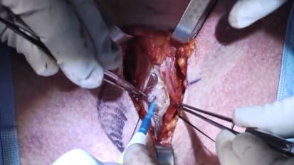
This is a right ear. The incision is marked approximately five to six centimeters behind the external auditory canal to allow adequate access to the mastoid for optimal surgical exposure. The incision is carried through skin and subcutaneous tissue and staggered slightly forward in the subperiosteal plane.
While this is based on surgeon preference, I like to grab a small piece of temporalis fascia to use later in order to block the mastoid antrum to reduce the risk of postoperative CSF leak. As you can see, a small piece of temporalis fascia is being harvested.
Next, using electrocautery, the staggered incision is made through the muscular periosteum to the mastoid cortex. The sternocleidomastoid insertion at the mastoid tip is rather adherent and electrocautery is often required to elevate the subperiosteum.
After the insertion of the sternocleidomastoid muscle is removed, the soft tissue elevates much more freely, and this can be performed with the subperiosteal elevator. The flap should be raised in the subperiosteal plane until the ear canal is encountered.
A wet raytec is then placed. And finally, fish hooks are used to retract the musculocutaneous flap forward.
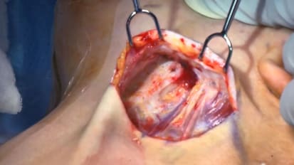
Next, a small cuff of muscle is obtained that will be used later to obliterate the middle ear space, again, to reduce the risk of post-operative CSF leak. Next, a wide cortical mastoidectomy with antrotomy is performed using the largest cutting drill bit possible with continuous irrigation.
Bone should be decompressed lateral to the temporal dura and posterior to the sigmoid sinus in order to gain adequate surgical exposure. Here, you can see the lateral semicircular canal. And in a minute, you will see the short process of the incus, both important landmarks for identifying the facial nerve.
After thinning the bone over the temporal dura, the thin layer of bone is decompressed to expose the temporal dura. This improves surgical exposure, improved hand working area, and illumination of the surgical field. In a similar way, bone over the sigmoid sinus is thinned.
And finally, the thin layer of bone is removed with a freer. It's important to perform this carefully in order to reduce your risk of accidental venotomy and significant bleeding. If significant bleeding is encountered from the sigmoid sinus, placement of gel foam, gentle pressure in some patients will stop virtually any sigmoid sinus bleed.
Bone in the sinodural angle is then removed. Superior petrosal sinus runs along this groove, and care should be taken to avoid injuring this.
Next, the labyrinthectomy is performed. This is generally first started by drilling the bone over the lateral semicircular canal. In order to protect the facial nerve at the second genu, the surgeon should not drill inferior to the inferior aspect of the lateral semicircular canal. Leaving this wallop as a buffer reduces your risk of inadvertent facial nerve injury during labyrinthectomy.
After drilling the lateral semicircular canal, the surgeon can drill more posterior, where they will encounter the posterior semicircular canal. And finally, the superior semicircular canal is located superior to both of these canals, and in a deeper plane.
The posterior semicircular canal and the superior semicircular canal meet at the common crus. Additionally, an artery can always be seen coursing through the subarcuate canal. This is another reliable landmark for the superior semicircular canal.
The confluence of the semicircular canals is at the vestibule, located essentially just deep to the second genu of the facial nerve. When drilling near the vestibule, it's very important to not undercut and accidentally injure the facial nerve as it courses through the tympanic segment.
Here, you can see the vestibule located just deep to the second genu of the facial nerve. The opening at the terminal end of the superior vestibular nerve can be seen in the vestibule. This is called Mike's dot. The floor of the vestibule marks the lateral extent of the internal auditory canal at the fundus.
The cochlear aqueduct can be encountered between the labyrinth and the jugular bulb. In small or medium-sized tumors, the cochlear aqueduct is often patent. And by opening it, egress of CSF can be achieved, which helps relaxation of the brain.
Next, the internal auditory canal is identified. It's important to realize that the lateral-most extent of the internal auditory canal is much closer to the surgeon than the medial extent at the porous. After the internal auditory canal is identified, superior and inferior troughs are performed such that at least 180, but more preferably closer to 270 degrees of the internal auditory canal is decompressed. Wide decompression is particularly important for large tumors that extend anterior to the porus acusticus.
The thinned bone over the internal auditory canal is finally removed, exposing the dura of the internal auditory canal. The medial segment of the facial nerve most commonly courses in an anterior superior location. Therefore, drilling on the superior trough, particularly near the fundus, must be performed very carefully to avoid injury to the facial nerve.
Next the posterior fossa dura is bipolar coagulated and opened. It's important to avoid arterial or venous injury when entering the dura, particularly if CSF could not be released earlier from the cochlear aqueduct.
Next, the dorsal portion of the tumor in the cerebellar pontine angle is stimulated with a prass probe. After confirming that the facial nerve is not in the unusual location of the dorsal pole of the tumor, the tumor is bipolar coagulated, the capsule is incised, and the tumor is internally debulked.
After the tumor is internally debulked, the capsule wall can be folded into the surgical field for removal.
After the posterior fossa component of the tumor is removed, the portion of tumor in the internal auditory canal can be removed. Again, the facial nerve is most commonly located in the anterior superior location within the internal auditory canal, and great care should be taken to avoid injury to the nerve in this location.
Here, you can see the tumor is being carefully dissected from the superior vestibular nerve as well as the facial nerve. The superior vestibular nerve can be cut early in the course of dissection, or some surgeons prefer to maintain its continuity during dissection, as it provides some additional support to the facial nerve and reduces the risk of stretch injury.
The facial nerve can be seen just deep to the superior vestibular nerve here. The remainder of the tumor is then removed.
After hemostasis of the posterior fossa is obtained, the middle ear is packed with muscle and fascia to reduce the risk of CSF leak. Some surgeons prefer to perform a facial recess with or without removal of the incus to directly visualize the eustachian tube for obliteration, while others indirectly obliterate this area through packing of the middle ear space through the antrum.
According to surgeon preference, an artificial neural substitute can be placed to reconstitute the posterior fossa dura. Fat is then placed in the mastoid defect.
The previously harvested fascia is then used to block the mastoid antrum. And finally, additional fat is used to fill the remaining portion of the mastoid cavity.
Bone wax can be used to obliterate any open mastoid air cells. Based upon surgeon preference, an absorbable mesh or titanium mesh cranioplasty can then be performed to secure the mastoid and fat in place and reduce the risk of pseudomeningocyle or CSF leak.
The incision is then closed in anatomical layers and a head wrap is applied. This concludes the surgical instructional video on translabyrinthine approach for vestibular schwannoma resection.