Chapters
Transcript
[MUSIC PLAYING]
BILLY BALLARD: Good morning. Would like to put in perspective the incidence of oral cancer, in oropharyngeal cancer. If we look at what's going on now, about 53,000 new cases of oral cancer will be diagnosed this year. And of those 53,000, 11,000 of those patients will die of that disease. And that's about 1 and 1/2 persons per hour per 24-hour day.
Now, these 53,000 patients, 65% of those will be cured of that disease and be alive and well five years from now. And we're seeing this tremendous advance and increase. And then the survival rate of patients over the decades. And this is true due to marvelous innovations in health care and early diagnosis.
And that's what we want to focus on today, looking at how do we recognize those reasons forever early and get them into treatment before this disease has advanced.
M ABRAHAM KURIAKOSE: Thank you, Dr. Ballard. So this is going to be a double act. Now, both of us are going to talk about the very important aspect of oral cancer. The outcome of oral cancer depends on the stage at which oral cancer is diagnosed. So one of the challenges we have is that on the whole, our patients tend to neglect oral cancer.
There is a study to show that there is about three months delay since the symptoms started before they seek help from one office in the community. And there is also data to show that there is about three months delay before a primary care provider suspect to diagnose cancer. So it took about six months' delay.
Now, the question is, how to compress that timeline. So we are going to discuss this aspect together and also purpose behind that. The reason is that the world in which I live-- I work in the cancer center-- the patients area already diagnosed with the cancer. So it's sort of of a no-brainer for me to do come up with what's the diagnosis waiting.
But the real world in which you live, it's different. You see a lot of patients with oral lesion, how to make sense out of these lesions. That's going to be the challenge. That's where Dr. Ballard comes in play, because he sees all of this every day, like all of you, so he can make sense out of these presentations.
Now, there are three objectives for this presentation. One is that how to distinguish a benign lesion from a precancer from a cancer but, equally important, how to separate that from a normal variation within the oral cavity.
And second, maybe very important, is that the tests assume that we have diagnosed a precancer lesion, how to start to define that this is a high-risk lesion. I need to send the patient to an oncologist immediately. And the last is just a general guideline. Once you suspect a lesion, what sort of flow we need to follow.
So that is a general kind of thinking, and I'm going to start with the conclusion or summary slide. I know some of you may fall asleep or something. But this is the most important slide of this 15 to 20-minute presentation.
Now, if you-- these are four categories which I was mentioning. That's a benign oral potentially malignant lesion, malignant lesion, and a normal variation. When a patient presents to us, we ask firstly history and then some examination finding.
In the history, we have to get three important points that can help us to make a diagnosis. First, is the duration. Second, is to whether there is any previous history of similar lesion. And third, pain, whether there's any associated pain on it. If the lesion is less than three weeks-- three weeks is very important-- less than three weeks, we suspect that it can be a benign lesion.
And if it lasts for months, then you consider this could be a potentially malignant issue. And malignant lesions on the whole should not heal within the two weeks' time. That two weeks' also is important. A normal variation, of course, we live with it. So it's been there for long time.
And previous history, that's important, because the benign lesion has not been present to previous similar type of lesions. Or a potential malignant and malignant not that normal variation of folks do not have that. But pain is another important feature. Most of the benign lesions will have pain, but malignant and premalignant lesions, the pain is not comparable or similar to the presentations. We see some other pictures.
And finally, the examinations. When it comes to examination, benign lesions, they have a very unique appearance. So we can learn from Dr. Ballard what is a benign lesion's appearance. And then when it comes to oral, potentially malignant lesion, there are three versions that it comes. One is the white patch, red patch, or mixed red and white patch.
Malignant lesions slightly similar to benign ulcer present as ulcer. Now, our job is to differentiate the malignant from a benign ulce3r. And that, we will come to that. Normal variation goes to the right and left side is the normal variation. Look and see one side. If it is on the other side, then we think that's a normal variation.
Let's get to the benign lesion side. It's a long list, a long list. I just put only about six of the common non-relation. Aphthous ulcer. Some of us get it. Traumatic ulcer. Some of us get it. And then denture hyperplasia, pyogenic granuloma. The broad spectrum of bullous lesions and Candida.
So let's see whether we can differentiate one from the other. So the next slide I'm going to put up here is some of the benign ulcers. I want Dr. Ballard to comment on what these are. These we see every day in our practice.
BILLY BALLARD: This, the first this you always see is the aphthous ulcer. And then aphthous ulcer, when we see, this is a benign lesion. Again, we deal with history. How long has the lesion been present? That is a superficial ulceration with this eosinophilic, this red border surrounding it.
This is a self-limiting lesion. So you want to know how long has it been present. Does it recur. And the patient will have-- and then the age of the patient. How old are you? What is the age of the patient? Usually, it's a lesion that we see in young individuals. And they recur repeatedly.
And the next lesion we're talking about is that of the traumatic ulcer. Now, with a traumatic ulcer, well again, history is important. How long has the lesion been present? Was there any incident that led up to the lesion? Is there a jagged tooth associated with that? Is there a recent, say, maybe a pizza burn or something like that? So all this is-- see, the history is again the things that we deal with.
The next lesion we see is that of-- and you see that the lesion has a purulent exudate that is associated with it. So again, it's a lesion that has healed. Then the next lesion we see is pemphigus. And here, we're talking about a chronic lesion that's been around for a long time.
And it begins-- and we'll talk about a bullous lesion. So the lesion begins really as a blister and it erupts and it goes over time. And then also want to see, are there other skin lesions associated? So all those things go together as we talk about this particular issue.
M ABRAHAM KURIAKOSE: Dr. Ballard, the duration, you said that's important. Is that right?
BILLY BALLARD: The duration, yes.
M ABRAHAM KURIAKOSE: So can you highlight on that, you know, how long. If an aphthous ulcer, how long it will last.
BILLY BALLARD: How long does it last? We see that most of the lesions should be gone in about two weeks. Now, this lesion, this is a chronic lesion and will go over long periods of time. And so that's what we're talking about here. And again, that would also have skin lesions associated with it as well.
M ABRAHAM KURIAKOSE: So pemphigus might last for a longer period but repeated on onset. So that's how that will give us an indication that's a benign lesion. There are three other lesions over here, Dr. Ballard, if you can just highlight.
BILLY BALLARD: So this is denture hyperplasia. We call it epulis fissuratum sometimes. And then this is especially where you usually have an ill-fitting denture that has rubbed and caused this. It's an irritative lesion.
And then the other one is the atrophic candidiasis. Again, this is a lesion that is inflammatory. We want to talk about this patient. What kind of history do they have? Again, this is especially when maybe recently treated with an immunosuppressant drug of some kind. And then the other thing is that we could actually just remove this and wipe that away. And this is something that would not happen with a lesion that is a true--
M ABRAHAM KURIAKOSE: See if I dropped-- Normally, we don't see these red patches. All right?
BILLY BALLARD: Right.
M ABRAHAM KURIAKOSE: We see this curdy white.
BILLY BALLARD: Right. Right.
M ABRAHAM KURIAKOSE: And you usually can--
BILLY BALLARD: Just sort of swipe it away, right. And then the peripheral giant cell lesion, again, this is a pedunculated lesion. It's sort of mass lesion. And this is a little bit more difficult. And you also have the red and the white and the leukoplakia as well as the erythroleukoplakia associated with that. So that's one that we probably would want to biopsy to make sure of what it is.
M ABRAHAM KURIAKOSE: So you don't sit on it. It's better to, even if you suspect clinically, better to do a biopsy. OK. Thank you. Then this is a very, very difficult problem we find in clinical practice. This white patch comes in the oral cavity.
The patient complains of burning sensation, everything. So main thing I need to differentiate, whether it's an oral premalignant lesion. Leukoplakia versus this entity called lichen planus. Can you tell us how do you do that?
BILLY BALLARD: Now, the lichen planus, as we see, one of the things as we look at this-- well, you see--
M ABRAHAM KURIAKOSE: The pointer doesn't work, yeah.
BILLY BALLARD: One of the areas here is--
M ABRAHAM KURIAKOSE: Doesn't work again.
BILLY BALLARD: It doesn't work. OK. Well, we're looking at the one on the tongue. The tongue is a white lesion, and it has a few areas of irregularity and sort of a lacy-type pattern. And then we look at the buccal mucosa. When we look at the buccal mucosa, again we see this lacy-like white streak. And that is the leuko stripe. And that's one of the things that we see as a characteristic of this lesion.
This is associated with an erosion. So this is an erosion lichen planus. And these lesions do have some kind of-- there was some discussion as to whether they actually are malignant. Are they inflammatory and really an immunological basis.
And again, we would need to do biopsy here because of the fact that we can wait if we are clinically confident of what it is, we may wait for a while. But still, we need to follow it up and probably biopsy it, because these do have a-- some of us believe that they do have some tendency to divide and propulgate into space.
M ABRAHAM KURIAKOSE: Very low, but about 3% risk of malignant transformation. The two erosive lichen planus.
BILLY BALLARD: Right.
M ABRAHAM KURIAKOSE: Another important feature for me to help me to differentiate is there is always a burning sensation.
BILLY BALLARD: Right.
M ABRAHAM KURIAKOSE: And leukoplakia does not have. Is that-- you said that?
BILLY BALLARD: That's true, yes. And also, these patients may also have skin lesions as well.
M ABRAHAM KURIAKOSE: Yeah. Skin lesions too. OK. OK. All right. That helps us to take you to the oral potentially malignant group of lesions. So if you can just show us-- I know the point unfortunately did not work. So if you can--
BILLY BALLARD: Now, this is a very initial in that it shows all of the stages that we want to talk about. We see the first one, the white lesion, the leukoplakia, the homogeneous leukoplakia on your left side. So that's one of the things we see. So that's the lesion that has very low incidence of malignant transformation, only about 6% of those will show maybe some malignant transformation or some dysplasia.
So as we move from that, we see the lesion that we have was the red and the white, where we call that erythroleukoplakia, a speckled leukoplakia. So we see those two things there. And with those two, is that that raises them to another level.
So at that case, all lesions have about a 50% chance of undergoing malignant transformation. And then as we move further, we see that's the smooth, thin mucosal surface. That is actually-- that's the erythroplakia. And has a very high incidence of undergoing malignant transformation.
And with that lesion, with that high incidence, it has about a 90% incidence of going here of undergoing malignant transformation. And then as we go further over, we see here, we see the lesion with the ulceration. And that is a true malignant lesion here, with ulceration.
So you've seen all the stages, from leukoplakia to erythroleukoplakia to erythroplakia to invasive squamous cell carcinoma. Now, with the invasive squamous cell carcinoma, not only do you have ulceration, but you also have the induration. So if you palpate that lesion, it would be a very, very hard, firm lesion, and that would let us know we're dealing with a malignancy.
M ABRAHAM KURIAKOSE: If I heard it correctly, as I looked at the color of the lesion, more red means bad.
BILLY BALLARD: That's right.
M ABRAHAM KURIAKOSE: That's good. OK. And then the white patch, if it is a homogeneous, I don't worry too much about it.
BILLY BALLARD: Right.
M ABRAHAM KURIAKOSE: OK. But all of the more or potentially malignant lesions changes the risk of malignant transformation, depending on the color of the new lesion or morphology of the lesion. OK, good.
BILLY BALLARD: OK.
M ABRAHAM KURIAKOSE: Now, this is a very difficult entity be facing our clinical practice, so-called proliferative verrucous leukoplakia. The term came to literature about 10, 15 years ago. Can you tell us about that?
BILLY BALLARD: Well, now this lesion, this is proliferative, a verrucous leukoplakia. And this lesion, again, it's very, very proliferative and it is growing very rapidly. It's a papillary lesion. And the lesion usually has a tendency, about 70% of these lesions will undergo malignant transformation.
But the good part is, I guess, is that they are fairly rare. But the rarity means that we may have a delay in the diagnosis. But again, this is a highly potentially malignant lesion. About 70% of these lesions will actually undergo malignant transformation. So the early diagnosis, early suspicion, and biopsy is very essential in this particular lesion.
M ABRAHAM KURIAKOSE: OK. Thank you, Dr. Ballard. And then one of the important things we see now-- once we see this patient there, how do I stratify them as a high risk on low risk? The reason is that malignant transformation just varies from about 3% to 30%. That's the literature says.
Now, how do we make a call? Without somebody sitting in the office, how do you make a call? Now, that depends on six factors. Six factors listed there. It's very important we learn that. One is duration. If the duration is more than two years, that's one of the red flags. Size more than 2 centimeter.
And then site is important. Tongue, floor of mouth is bad risk, high risk lesion. Then so-called nonhomogeneous leukoplakia, which Dr. Ballard mentioned. And then patient presents without-- I repeat-- without a risk factor, that is a bad prognostic factor. And also a presence of high-grade dysplasia.
So if you have a patient with those two pictures, one on the buccal mucosa, another one on the that buccal mucosa with that erosive kind of patch, that's the erosive ones which we call it at a high risk. So that helps us to differentiate high risk versus low risk.
Now, next category of the lesion is the malignant lesion. Now, Dr. Ballard, you know, cancer is a rare problem. But we see a lot of lesions in the oral cavity. Where should I look inside the mouth to find the cancer? Cancer happens all over the mouth? Any particular area?
BILLY BALLARD: Well, the highest-risk areas, not a part of the tongue, ventral surface of the tongue, floor of the mouth. Those are the lesions-- those are the areas where you showed very, very high degree of suspicion of lesions that occur in that area. And those areas with ulceration, the duration of the lesion and whether or not the lesion is ulcerated, as we see in the lesion here, and if the lesion is firm to palpation. So those are the places that we see in malignant lesions.
M ABRAHAM KURIAKOSE: So look for the common sites, tongue, floor of mouth, and the sulci of the oral cavity. This is a buccal mucosa alveolar lesions. And then how do I differentiate-- you mentioned about the duration, then the pain is you don't have the proportional--
BILLY BALLARD: The pain isn't-- right.
M ABRAHAM KURIAKOSE: And then any clinical features I need to elicit during the examination?
BILLY BALLARD: It's ulceration and induration. They're a very, very firm, hard lesion. They say stony hard lesions are one of the things that we see with malignant lesions.
M ABRAHAM KURIAKOSE: OK. Quickly go through some other normal variation--
BILLY BALLARD: Some of the normal variations we see is Fordyce granules. Those are ectopic sebaceous glands. We see those on oral mucosa many, many times. So this is a developmental lesion that has no real significance other than to recognize it and assure your patient that everything is going to be OK.
Another thing is the lesions that are on the tongue or the hyperplastic folate papillae. That is another thing that we see. Another one is here the circumvallate papillae that we see in the posterior area of the cavity. So those are things that we see that normally, and we can-- when the patients come in, we can recognize those and ensure them that those things are benign and of no consequences.
Here again, is a fissured tongue. And this again is a developmental abnormality. And the hairy tongue. And that's a hyperplasia of the villae on the tongue. And many times, they will pick up various colors. They may be pink if you've been drinking wine. They may be purple or whatever, but they have those kinds of colors that we see.
The other thing is, the other lesion we see is the geographic tongue or the-- and we also call that migratory glossitis. And that's a thinning of the surface epithelium. No consequences. And those lesions, they said geographic because of the figures they make on the tongue.
And then also migratory, because they will go from area to area. So again, those are the kind of things we want to talk to our patient about and share a-- and get a history.
M ABRAHAM KURIAKOSE: OK. Thank you. The last two slides, we just want to show what should be the workflow when you see your patients in your clinic. The first thing is that, let's say that the patient is presenting with a white or red patch. We mentioned about that six high-risk features, we look for them. Site, size, and other features. And then we look at the history.
So from that, we can say that this is a neoplastic lesion or a nonneoplastic lesion, so-called benign lesions. Benign lesions, we can follow up. And if it a potentially malignant or malignant lesions, we have to stratify them into a high risk to low risk based on the six features which I mentioned. If it is a high risk, don't wait. Go get a biopsy done. If the low risk, we have time.
So we remove the irritant factors like sharp teeth and so on and wait for two weeks' time. Now, this is important-- two weeks time. If it resolves, then we don't have to worry too much about it.
But we have to keep the patients on follow up. If the lesion persists, we have to do a biopsy. And once we have done the biopsy, we get two answers. One is that where there is a nondysplastic or mild dysplastic lesion, that we can stratify into the group of low-risk lesion.
Again, we need to keep an eye on the patient but not that stringently. But the high-risk lesion, it has got a very high risk of malignant transformation. That is a high-- moderate to high-grade dysplasia. Most of time, we do excision. Very controversial. But I'm sure you'll come to that during the panel discussion. And then we need to follow up the patient on the long term.
So finally, the summary slide. So we learned three things. One, we learned about that benign versus potentially malignant to malignant lesions, and we looked at some of the normal variations and that six features to separate the high risk versus low risk we learned.
And finally, one important take-home message, if you don't remember anything during this morning session, if an ulcer persists in the oral cavity, which lasts more than three weeks, time to get a biopsy done. Thank you.
[MUSIC PLAYING]
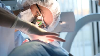
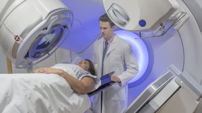
Billy Ballard, DDS, MD, and M. Abraham Kuriakose, MD, put into perspective the essence of oral cancer. At the current rate 53,000 new cases of oral cancer will be diagnosed this year and of that 11,000 of those patients will die from that disease. This lecture covers the advances in diagnosis and treatments to increase the rate of survival.
Related Presenters
Postdoctoral Fellowship: Roswell Park Memorial Institute
Billy Ballard, MD, DDS, is the Associate Dean for Continuing Medical Education; Chairman and Professor, Pathology, Meharry Medical College. His research specialty is Anatomic/Surgical Pathology; Head and Neck, breast, genitourinary (prostate), ...
Director, Translational Research for Head & Neck / Plastic & Reconstructive Surgery
Moni Abraham Kuriakos, MD, FDSRCS, FFDRCS, FRCS Ed, FRCS, BDS, is the Director, Translational Research for Head & Neck / Plastic & Reconstructive Surgery at the Roswell Park Comprehensive Cancer Center.
Related Videos