Nobody has a crystal ball that will accurately predict exactly what someone’s prostate cancer will do – but Hopkins pathologists are working on it.
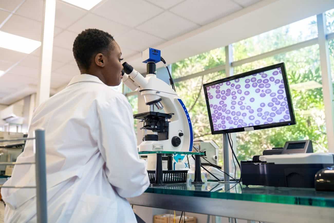
This crystal ball involves a computer: After a prostate biopsy, the pathologist takes the tissue cores and puts them on a glass slide. The pathologist analyzes them; so does a computer – and “this is revolutionizing the way pathologists diagnose and grade malignancies,” says pathologist Tamara Lotan, M.D.
Already, artificial intelligence (AI) and machine-learning algorithms are helping to improve the precision of Gleason grading, to standardize it “and remove human subjectivity from the process.” Taking this to the next level, Lotan and pathologist Angelo De Marzo, M.D., Ph.D., are endeavoring to teach AI, and “herein lies the future of digital pathology,” she says.
Can AI learn how to identify molecular characteristics in prostate cancer cells more accurately than the human eye, and to predict the patient’s course of disease? Lotan and De Marzo believe it can. This year, funded in part by the Prostate Cancer Foundation and private philanthropy, the scientists and colleagues teamed up with AIRA Matrix, a company that uses “deep learning” (involving layers of neural networks).
“We trained deep learning algorithms to identify prostate tumors with two of the most common molecular alterations, ERG gene rearrangements, and PTEN gene deletions,” says Lotan. “By eye, we can’t just look at a prostate tumor and tell whether it has these underlying genomic alterations.” But with training, “we showed that the computer had excellent accuracy in predicting which tumors had these genetic changes.
Lotan and De Marzo hope this will “pave the way to inexpensive and accessible screening tools,” to determine which patients need additional genetic sequencing and to guide precision treatment of advanced cancer. Their work was recently published in Modern Pathology.

