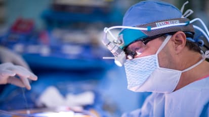
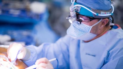
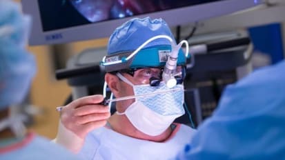
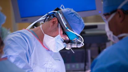
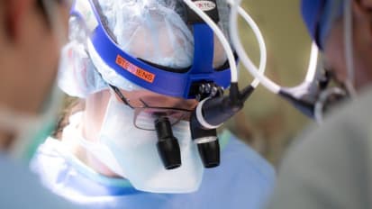
Mayo Clinic otolaryngologist Eric J. Moore, M.D., demonstrates a total parotidectomy performed for cutaneous squamous cell carcinoma metastasized to the parotid gland.
Eric J. Moore, M.D.
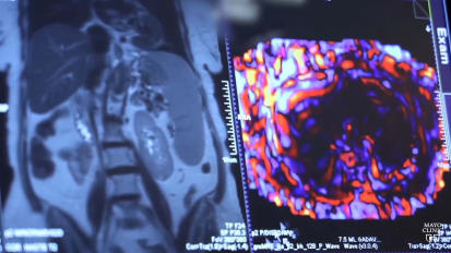
Hi, my name is Dan Price. We'll be doing a total laryngectomy today. This is a patient with A T four larynx cancer. Never had any prior treatment. Preoperative evaluation includes office and operative endoscopy. To understand the extent of the tumor and the extent of resection required. Patient has extra laryngeal extension as can be seen here on the C T scan is not a candidate for laryngeal preservation surgery or therapy. We try to make a separate neck incision from the stoma and have at least three centimeters of skin between these to preserve the blood supply to the skin between the neck incision and the stoma. This decreases the rate of visualization flaps are elevated for the next section. The hyoid bone is identified and palpated. We divide all of the strap muscles. There's really no utility to preserving strap muscle for closure or reconstruction and dividing these as close to their origins and insertions is helpful. Sternal hyoid and sternal thyroid muscles are divided inferiorly with a subglottic tumor or extra laryngeal extension, least hemi thyroidectomy on the side of the tumor should be performed. One has to use caution not to injure the 12th cranial nerves. So the hypoglossal nerve should be identified on both sides. The vascular pentacles should be divided superiorly. This process is repeated on both sides. The homo muscles are divided inferiorly. The ph constrictor should be divided off of the thyroid cartilage. You can preserve some of this musculature for your closure as well as some of the perio if there's not tumor involvement in these regions. So I will dissect down to the lateral aspect of the thyroid cartilage and then dissect in a subpar plane superiorly. The P form mucosa is surprisingly close to that superior thyroid corne. And so taking some extra time to not create a phony here is worthwhile as you'll have to resect those areas if you create omy, otherwise you increase your risk of official. So here, the inner peron is bluntly dissected off of the thyroid cartilage on the opposite side of the tumor because there's a significant amount of tumor on the contralateral side. A lot of this work will be done after we've visualized the tumor on the side of the tumor at this junction, we perform the tracheotomy, no Bjork flap should be performed. It's important to try to avoid performing a tracheotomy prior to surgery. As this increases the risk of recurrence. We've performed laryngoscopy and know the extent of the tumor inferiorly and perform the tracheotomy as high as possible. Achieving a adequate surgical margin. This gets the endotracheal tube out of the way we now dissect down to the hyoid bone and release the super hyoid musculature. Again, awareness of the location of the hypoglossal nerves uh should be kept in mind here. Similar to the thyroid cornu. When you take out the corner of the hyoid bone, it can be tucked into the phom mucosa and you can make an incidental pharyngotomy. If you want to stay on the exterior surface or lateral surface of the hyoid bone, as you dissect it out and then as you work on the deep surface of the hyoid bone stay in very close proximity to the bone. Here, you can see the shiny high optic ligament. So we divide the hyoglossus muscle and enter into the molecular, the mucosa of the molecular contralateral to the tumor is entered. So that cuts around the tumor can be made under direct visualization. You can see inserting the digit into the pure form sinus and then under direct visualization, cutting along the high epiglottic ligament preserving as much of the normal mucosa as possible, which will allow you to have a linear vagotomy closure at the end of the procedure between my index finger and thumb is the tumor as it extends out laterally. And these pharyngeal mucosa cuts are made under direct visualization, maintaining as many centimeters as possible around the tumor. You can see tumor in the post cricoid area here as well and that mucosa should be resected. We then need to enter into the partition between the trachea and the esophagus. This can usually be done bluntly. Again, most of these, the post cricoid mucosa is normal. So under direct visualization, we sharply divide that so that we maintain as much mucosa as is oncologically responsible to make for a tension free watertight closure, keeping my finger in the esophagus as we then elevate off the partition between the membranous trachea and the esophagus delivering the laryngeal specimen. The specimen is inspected for any close margins. Margins are taken off the primary specimen, Acular subotic margin examined. The stoma is then fashioned again a circular stoma about two centimeters in diameter preserving 2 to 3 centimeters of skin between so that skin is well vascularized. The stone has matured with absorbable sutures here, I'm using vil. Although I'll often use chromic sutures as I find the vico sutures stay too long. The patients get small abscesses around these and chromic are perfectly adequate. The suturing is done to cover the edges of cartilage with a vertical mattress suture, bringing the suture around a ring of the trachea, just cure it. This will help prevent infection as well as stenosis phom myotomy is performed inserting a digit into the esophagus. The phal muscle can be easily felt here, cut it sharply preserving the esophageal mucosa, avoiding a full thickness defect. You can see that there's still constriction at the inferior aspect of my digit. You have to get this entire muscle. The patient may have speech problems supposed to operatively that optimizes their tracheoesophageal speech. Ideally, the phony is closed over a feeding tube in a linear fashion, avoiding a T closure. As the trifurcation is an area of weakness. Most likely to break down. You want to invert the mucosa. This can be done with a variety of stitches. Here we do a stitch that is a running inverted stitch. You go through the muscle edge and out through the muscle and then cross over to the other side, going through the muscle edge to invert and not have mucosa in your closure, which will increase your risk of a leak as you reach the most cephalad aspect, you then include the mucosa of the tongue base in the molecular. Whenever possible, I will try to do a second layer of closure using the phal constrictor muscles to bolster your mucosal closure if the patient has had prior irradiation and they should get a peck flap. And this has been shown to decrease the rate of official by nearly half skin closure is then done in the standard fashion suction drains are placed. Patient is admitted for laryngectomy care and teaching postoperatively will perform a swallow study about 7 to 10 days in a primary patient. Key points for total laryngectomy include performing endoscopy preoperatively or inter operatively to understand the extent of tumor, doing a separate stomal incision from the neck dissection incision creates a better stoma with less risk of salivary fistula, preserving as much normal mucosa as possible allows for a tension free water tight closure and decreases your risks of a fistula. Complete cricopharyngeal myotomy should be performed to maximize the patient's tracheoesophageal speech. And patients who've had prior chemo radiation therapy should have pectoralis flap or other free tissue coverage to decrease the risk of visualization. Thank you for watching.