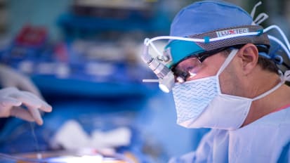
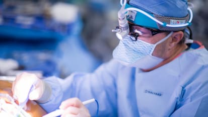
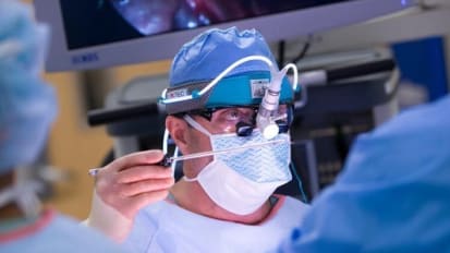
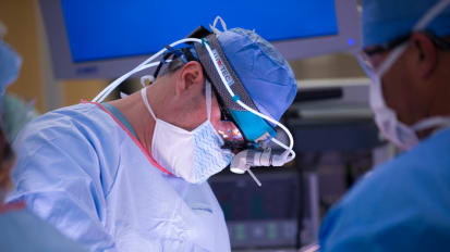
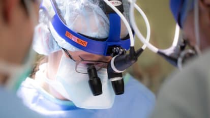
Mayo Clinic otolaryngologists Janalee K. Stokken, M.D., and Christopher M. Low, M.D., demonstrate an endoscopic orbital decompression in a patient with Graves' ophthalmopathy.
Janalee K. Stokken, M.D.
Christopher M. Low, M.D.
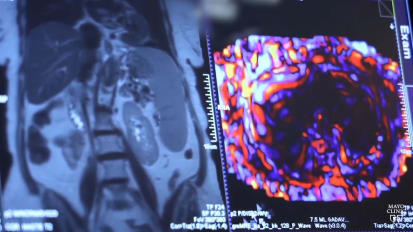
This is Christopher Lowe presenting a case of endoscopic oral decompression with Doctor Jana Lee stoke. The most commonly performed indications for endoscopic oral decompression are for functional or cosmetics of thyroid eye disease. Also known as graves ophthalmology. Functional aspects include restrictive myopathy leading to Tilia exposure, Caratti or optic neuropathy. Cosmetic considerations include an altered appearance secondary to proto and opthalmic. Other indications for endoscopic orbital decompression are for access to the orbit for the removal of benign or malignant orbital tumors. Biopsy of indeterminate lesion, decompression of Orla abscesses and hematoma. And as an approach for optic nerve decompression. This patient is a patient with opus secondary to thyroid disease. Endoscopic oral decompression is performed under general anesthesia. As an outpatient procedure, it first starts with a max intros toy total ethmoidectomy with skeleton organization of the lamina PAA and anterior skull base depending on a patient's anatomy. A frontal sinusotomy is often performed and consideration can be given to performing a sphenoid atomy with the goal of preventing postoperative postobstructive sinus disease, secondary to prolapse oral contents in the sno nasal cavity. Further details about endoscopic sinus surgery can be found in other videos in the surgical video atlas. Once wide exposure has been established, mucosa is first removed from the lamin of paprika. Next, the bony lamina is meticulously removed without fracturing and cutting instruments, carefully preserving the periorbita and skull base. The para orbita is kept intact while removing the Laina to keep orbital fat from obstructing the surgeon's view. During dissection with the periorbita exposed, an initial posterior periorbita cut is made in an inferior to superior direction. The choice of instrument is as per surgeon preference. Here, an arachnoid knife is used, care is taken to ensure only the pa orbiters in size by seeing the tip of the blade through the tissue. During the cut. Following this, a superior cut is made in a posterior to an interior direction. Here, an arthur luck retrograde beaver blade is used and care is taken to keep the leading edge of the instrument visible through the periorbita. An inferior cut is made from a posterior to interior direction and the periorbita is peeled away using grasping forceps, retropulsion of the eye helps to identify fascial bands in the oral of fat which are then divided with blunt or sharp instrumentation. The facial band cuts are made in the same orientation as the medial rectus to avoid injury to the muscle fibers underneath. As the fashion bands are divided, the globe feels soft into palpation