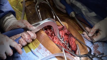
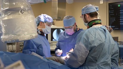
Neurosurgeon Stephen Kalhorn, M.D. , uses a 3D digital exoscope to perform a minimally invasive tubular microdiscectomy to relieve pain in a patient with a herniated disc. He explains the transformative power of this new tool, which gives high-definition global views in a more relaxed and comfortable manner than that achieved with a conventional microscope. This creates an easier, more streamlined experience for patients and surgeons and gives the surgical team the capacity to work together synergistically by observing the same detailed view.
so the patient was a gentleman who had horrible leg pain. Some people call that Siderca. It's pain that originates from a nerve in the lower back that radiates down one or both legs. The patient had a big lumbar disc, our nation. On his MRI scan. We had sent them for a variety of non operative modalities to try to get him better without having to do surgery. He got steroid injections, went through physical therapy and all the routine conservative things to deal with this type of problem. So despite that he still had significant pain down the leg that was quite severe and interfering with his ability to enjoy his life. The procedure that we offered him was a minimally invasive or tubular micro step to me, where you make a small, roughly 18 millimeter incision on the patient's back and Doc, a series of dilator is down on top of the lamb eater bill, which is the bone just next to the nerve words in the low back. After you've docked down with one of these dilator is you can place a minimally invasive retractor system which looks like a small tube which is attached to the bed. At that point, you drill away some of the bone, remove what's called the ligament, um, on top of the nerve roots there, and gently retract the nerve root over and take out the hard disk. If you're used to using the microscope, there's a little bit of a transition period to get used to using the three D exit scope. You're wearing three D glasses, and your posture just needs to be relaxed from the beginning. So when you start using the exact scope, you just need to realize that you can relax. You can use your muscles in a more ergonomic fashion with the exact scope technology you're able to more easily and comfortably take out a discrimination and someone's back, especially through a minimally invasive surgery, which traditionally has been limited by the viewing ability through the operative microscope. The exact scope allows you to zoom in with with higher definition than the regular microscope. In my opinion, you're able to see different angles that you necessarily would have a lot of different. A lot of trouble scene with the traditional microscope. The you're able to easily move the exit scope around in different angles, in your operative field with a robotic arm attached to the exact scope. You're able to put a pointer down into the field and point exactly where you want to see, and the endoscope will move into that line of sight. The exact scope will store different positions, so you can say that you want to look more immediately or laterally, and the endoscope will move into that position. There was a store settings and go to that. Go back to that initial position later on the surgery so you can flip back to that other view and not having to remember where that was, or you have to maneuver a big microscope around in that field. Well, I think it just overall reduces a lot of or frustrations. And so I think if you're able to provide a procedure where that's easier and more comfortable and fluid for your patient than the patients are going to have a less time under general anesthesia and the easier time getting through their surgery, it just makes it also a lot easier for everyone in the room to see the surgery for the your scrub nurse and the circulator to anticipate what needs to happen next. You find that your scrub nurse is able to see what the next steps are on the surgery, and he or she will just start handing instruments without having to ask for them because they know the upcoming steps. That's that's not always possible with the traditional microscope. So it really builds a team environment in the operating room. It allows you to more efficiently and safely take care of the patient.
Related Presenters