Bernard R. Bendok, M.D. , Harry Cloft, M.D., Ph.D. , Kelly D. Flemming, M.D. , and E. Paul Lindell, M.D. , discuss how Mayo Clinic physicians use advanced imaging software, augmented and virtual reality, and a multidisciplinary team to diagnose and treat arteriovenous malformations (AVMs).
Physicians prep for surgery using advanced simulations that help minimize surprises that can appear during a surgical procedure. After, the team creates an individualized treatment plan that puts the needs of the patient first.
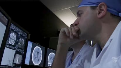
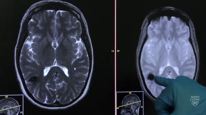
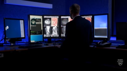
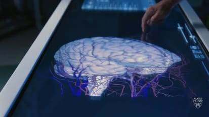
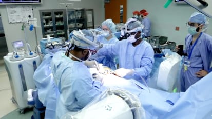
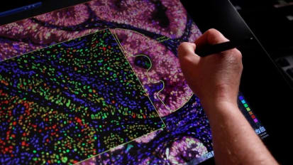
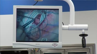
here at Mayo Clinic. We have an A. V. M. Conference where we talk about a VMS as a group and we try to focus the care amongst a small group of individuals who have developed tremendous expertise. Our radiologists, for example, are extremely experienced with picking up subtle signs of hemorrhage on an M. R. I. Or a cat scan and then looking for subtle signs of an A. VM. On a scam that may not be obvious on a superficial review. It's very characteristic appearance in an A. V. M. Where there's a tangle of blood vessels there that shouldn't be there. The combination of CT angiography with advanced NMR techniques that can look at the structure of the brain, the structure of the AvM and even the function of the brain tissue around the A. V. M. Then a multidisciplinary team would look at that angiogram and make decisions about what is the best treatment. The treatment of A VMS has gone from a primarily surgical approach to a combined endovascular approach. Usually it's open surgery or radiosurgery that ultimately cures that a VM but sometimes blocking off some of the blood supply through a catheter can help those procedures be safer and more effective when a patient presents with hemorrhage, it's very important to have a very advanced critical care team that can manage the patient medically to reduce brain pressure and in some cases we can go in and seal the rupture site with glue or coils and then we bring the patient back down the road once they've recovered to definitively treat the A. V. M. And that requires the expertise of a neurosurgeon, a vascular neurologist, a radiation oncologist who all specialize in treating a VMS. We've recently added augmented and virtual reality to our platform so that we can study are a VMS in three dimensions using three D prints, but also virtual reality platforms. And then we take that data set, that knowledge that we've gained. So we can together with our teams are nurses and technicians rehearsed the operation so there are no surprises or we minimize the chance for surprise because we can simulate things that can go wrong to make us better practitioners. We continue to get a lot of patients referred to us that provides us with continuing evolution of experience so that we can come up with good diagnoses and therapies. And also the basic principle that we have a patient comes first and offering compassion I think is extremely important.
Related Presenters