Volume 90%
Press shift question mark to access a list of keyboard shortcuts
Keyboard Shortcuts
Play/PauseSPACE
Increase Volume
Decrease Volume
Seek Forward
Seek Backward
Captions On/Offc
Fullscreen/Exit Fullscreenf
Mute/Unmutem
Seek %0-9
00:00
00:00
00:00
 

Transcript


SPEAKER 1: The first case for Dr. Zhou is going to be a per oral endoscopic myotomy for type II versus type III achalasia with EndoFLIP 2.0. Briefly, this is a 61-year-old woman with a history of only anxiety and depression presenting for the management of her achalasia. Her duration of symptoms have been two years. Her Eckardt score is 9, consistent with weight loss of less than 5 kilograms, dysphagia with each meal, retrosternal pain daily, and regurgitation with each meal.

Here we see the esophogram consistent with stage I. The measurements are 2.1 centimeters, and it's in a nonsigmoid configuration. Here on the manometry, we see panesophageal pressurization, with areas of intense spasm. The basal LES pressure is 101. EndoFLIP 2.0 will be used to determine whether this is type II versus type III achalasia, which will help curtail the length of the myotomy that needs to be performed.

STAVROS N. STAVROPOULOS: We're in Dr. Zhou's room, because he has the case that there is a debate about whether it's a type II or type III patient, and where EndoFLIP may make a difference. So I'm here doing the EndoFLIP before he starts. The point I'm trying to figure out, if we are going to do a short myotomy or a long myotomy.

So you can see, for example-- let me show you. Let me pause here. OK. So let me show you what the EndoFLIP sees. So this is the EndoFLIP 2.0, which apart from a fixed image of the balloon shape, it gives you a continuous tracing. You can see as it's moving with time, when I pause it now to discuss the finding.

So the balloon has 16 sensors that detect impedance, that conducted small electrical currents and detect how much hypertonic saline is around them in the balloon. And that allows them to plot the shape of the balloon. And then out of that, we can calculate all kinds of things, such as the tightest diameter, the cross-sectional area, and then knowing how much saline you have in the balloon, and you can pick 30, 40, 50 mLs. Knowing the volume and the pressure of the balloon-- there's a pressure sensor-- you can calculate things like compliance and distensibility.

The most useful number is distensibility, which divides cross-sectional area and pressure. So if you have a very large cross-sectional layer with very low pressure, that's a very high distensibility. It's a very open sphincter. Whereas, if the cross-sectional layer is very small and the pressure in the balloon is very high, that's a very tight sphincter.

Untreated achalasia has distensibilities in the 0.5 range. Properly treated achalasia, whether with POEM, Heller, or something else, should have distensibilities of 3 or more. And therefore, it's very usual. Her distensibility right now 0.6 at 30 mLs of saline. So this is very consistent with very tight virgin achalasia.

Now question number two-- and this often comes handy when you have an equivocal manometry where the patient walks, talks, looks like achalasia, but for some reason the IRP is not perfect nonrelaxing-- if you do an EndoFLIP, and you get a distensibility of 0.6, you can actually clarify that this is indeed achalasia.

Another area of unclear manometric diagnosis is, what's type II and what's type III? Very low interobserver agreement, even among experts sometimes. And here it's very important for treatment, because for type III, you need a long cut, for type II, you need a short cut.

So this is such a situation here. The manometry was read as a type II versus type III, because of this spasm in the distal esophagus. Let me run the EndoFLIP. But you can see, normally the sphincter is about 3 centimeters or so. Here you get a much longer spastic segment. What you want to see is a red sphincter with blue on either side.

Here, you see the sphincter. You also see some movement here. There is a contractility. Repeatedly we demonstrated some persistent spasm extending from the sphincter proximally. So you can see, you're just seeing a contraction right here. You see that? There's a retrograde contraction that just took the entire distal part of the esophagus. Let me pause it there.

CHRISTOPHER J. GOSTOUT: Yeah, we can see that pretty nicely.

STAVROS N. STAVROPOULOS: I don't have any way to point for you. But the first half of the tracing shows a stable red zone, which is a long, tight sphincter. And suddenly, this red zone expanded to include several centimeters of the distal esophagus. And this is that distal spasm.

And then you can also see a separate red line at the top of the tracing, which appears to be another area of isolated spasm, another about 10 centimeters proximal to the sphincter.

Now, the typical findings of a type III patient would be retrograde contractions. You would see those waves again, and again, and again, like snake tracks in the sand, going left to right upwards, instead of the usual contractions, which is left to right going down. And these are called "retrograde contractions." That's a typical finding in type III.

We haven't been able to see this here. So I think from the EndoFLIP perspective, there may be an area of distal spasm, but it doesn't have the typical type III findings that we've seen with the EndoFLIP in type III patients. So I think here, we have to do some compromise myotomy-- maybe not quite as short as a type II, and not as long as a typical type III. I would include the sphincter maybe a few more centimeters.

And Dr. Zhou can use his vast experience of thousands of POEMs to also do an endoscopic assessment of where the proximal length of the spasm is, and where he should start his myotomy.

PING HONG ZHOU: Yes. We must check the esophagus with the scope to determine where--

STAVROS N. STAVROPOULOS: Where the scope-- and to see where the spasm starts. But then in the last few years, I favor shorter rather than longer, because ablating even a type III patient's motility aggressively is not a good thing, I think. Even for type III patients. So generally, we have all shortened our myotomies.

Another trick I use is sometimes I do a long tunnel, which doesn't really commit the patient to a long myotomy. Then I do a little test myotomy at the top of the tunnel. And if the test myotomy reveals a muscle, like a centimeter thick or something, or 7 millimeters, that goes with the type III or a spastic disorder. Then you do a long myotomy.

If the test myotomy a very thin muscle, you cannot do a long myotomy, and go down to near the GE junction and do a short myotomy. So I can do sometimes a long tunnel, do a little test, and see how thick the muscle is.

PING HONG ZHOU: We perform a US to check the muscle--

STAVROS N. STAVROPOULOS: With US is another way of doing it. Right. Yeah, we can do a US.

PING HONG ZHOU: We have to see the thickness of the muscle.

STAVROS N. STAVROPOULOS: I don't have as much experience judging what's thick only US, as I do on cutting a little muscle. And I think the thickness on the US is also affected by how much distension the esophagus has. So it may look thinner or thicker, depending on how much distended it is. But if you do a little myotomy, we all know what's thick and what's not thick, from experience.

So sometimes I'll do that. I'm not sure we'd need to do this here. Maybe do just a longer type II kind of myotomy.

CHRISTOPHER J. GOSTOUT: Stavros, can I interrupt you? Let's go back to the EndoFLIP. So above the intense red area, you see that's coming on top of that, there's a little band on top, right? It's got some red. So that indicates maybe the myotomy should go up to that level.

So my question is, can you accurately measure the length of your myotomy with the EndoFLIP? Can you say, OK, the bottom is going to be here, and the top is going to be here. That matches that slight red band on top.

STAVROS N. STAVROPOULOS: Yes, because--

CHRISTOPHER J. GOSTOUT: Or is it just an estimation when you finally get in there with the endoscope?

STAVROS N. STAVROPOULOS: You can measure very accurately with the EndoFLIP, because you see the white lines, I think, maybe in the balloon representation. You can see, you just saw a-- see? The patient just went into complete spasm of the distal esophagus. You see that?

CHRISTOPHER J. GOSTOUT: Right. It's used up the entire image. It's all redded out.

STAVROS N. STAVROPOULOS: So you can tell where the top spasm is by using those demarcations of the transducers. We calculated it to potentially having a area of fixed spasm approximately 10 to 12 centimeters from the beginning of the sphincter area. It's a high pressure zone.

PING HONG ZHOU: And we determine length of the myotomy based on the EndoFLIP.

STAVROS N. STAVROPOULOS: Right. The EndoFLIP helps. In this case, I can say it's classic for type III, because then it helps a lot. Then you do a long myotomy. But here you can see, she just went into distal spasm.

The problem is, I've seen this a lot with type II patients that have good pressurization waves. And I think this may help their emptying. So I used to be more aggressive in considering these patients, potential type III ones. See hear, I'm running it again, and the spasm persists. It has persisted now as long as I've been talking.

And now it got even tighter. You see the top? It's like obliterated completely the lumen. So this is now running in real time. You can see this spasm now persists.

CHRISTOPHER J. GOSTOUT: Right. And it's actually more intense on the top.

STAVROS N. STAVROPOULOS: Yeah, it's really very impressive, I have to say. See, now it relaxed. You see? But I think this could all be consistent with a very tight pressurization type II. So I don't think we should treat it as a type III. But Dr. Zhou will also assess endoscopically the areas of spasm.

CHRISTOPHER J. GOSTOUT: So when you tell us you're going to do a shorter myotomy, or it's trending towards shorter myotomies, and this one is going to be maybe a little longer as opposed to a long myotomy-- give us some numbers. What's short? What's a little bit longer?

STAVROS N. STAVROPOULOS: I'll give you my numbers, because Dr. Zhou may have different numbers. But I've now settled-- because we all went from short to long, and now we're all shortening again. I'm down to maybe 5, 6 centimeters for a typical type II.

PING HONG ZHOU: Type II?

STAVROS N. STAVROPOULOS: Well, no, actually type I is definitely 5 centimeters. Type II may be 6 centimeters, 7. And for type III, I'm down to maybe 15. When I have done myotomies as long as 20 to 26 in years past.

CHRISTOPHER J. GOSTOUT: Does this include the gastric side? So what's your gastric side? What's your esophageal side?

STAVROS N. STAVROPOULOS: Gastric sides, I believe we are both shorter than 2. We used to do maybe 2 or 3 centimeters on the gastric. Now we maybe are 1 to 2. I don't know, Dr. Zhou, what's your numbers?

PING HONG ZHOU: 2 or 3.

STAVROS N. STAVROPOULOS: For type I. What length myotomy type I, what length for type II, what length for type III?

PING HONG ZHOU: In Shanghai, we base it on the manometry and the endoscopic findings. So where is the spastic region. And then we just make a tunnel proximal to the spastic region, to make a tunnel. But even for the sigmoid type, always make a short tunnel, for sigmoid type.

STAVROS N. STAVROPOULOS: So what's your average myotomy for type I?

PING HONG ZHOU: Type I, I think very short-- 7 or 6, 6 or 7.

STAVROS N. STAVROPOULOS: Yeah, I'm more like 5 for type I. And for type II? You're maybe 8, 9?

PING HONG ZHOU: Always, the typical, I think, traditional POEM is 10 centimeters.

STAVROS N. STAVROPOULOS: Right. He's more like 9, 10 for type II, and 7 for type I. I'm about 2 centimeters shorter on those. And for type III, as he said, you adjust it, depending on where you see the spasm on manometry, on EndoFLIP, and on endoscope. So type III is very variable. Much more variable.

PING HONG ZHOU: 15, 20, or 25.

STAVROS N. STAVROPOULOS: Right. It could be as short as, we know, 15, and as long as 20, or 21, or 25.

All right. So I'm going to go to my room to start my POEM, because we're falling a little behind. And I'll pull everything out, and Dr. Zhou can start his over here.

PING HONG ZHOU: So first we remove the balloon.

STAVROS N. STAVROPOULOS: We deflate the balloon.

PING HONG ZHOU: And then we remove the balloon. And then--

STAVROS N. STAVROPOULOS: Ronnie, can you help? I've got to go.

CHRISTOPHER J. GOSTOUT: For the audience, remember you can text in questions as these cases are moving along. And we'll interrupt and answer your questions as they come.

PING HONG ZHOU: And now we're moving the balloon off the EndoFLIP from the patient's esophagus. And now I begin to check the esophagus with the scope. The CO2 is OK? CO2.

CHRISTOPHER J. GOSTOUT: So as Dr. Zhou is preparing his site for his tunnel, getting back to the EndoFLIP, it's a tool you can also use after the procedure to check on your results to see if whether or not it's satisfactory. And especially in this indeterminate case, whether or not your myotomy was long enough to cover that more proximal area where we saw that intense spasm.

PING HONG ZHOU: So before the POEM procedure, the esophagus preparation is very, very important. From the endoscopy, we can now see the lumen is dilated. So I flush the esophagus with the saline. Here is the stricture area. So you can see here this junction is very, very tight.

And here, the length for this patient is 36 to 37 centimeters from patient's mouth. I push the scope forward. Here is the EG junction, I think. So now the scope passes through the EG junction. So I feel tight of the EG junction. Now I will flex the scope. So we can see the scope was wrapped by the muscle.

So this is the EG junction. And the length of the scope, for this patient the EG junction is about 36 from the patient's mouth. So now I pull back the scope. So this is the dilated lumen. I think here is the spastic region. Now the scope is 26 centimeters. So that means 10 centimeters proximal to the EG junction.

10 centimeters from here, I began to make a tunnel. Do you agree with me?

CHRISTOPHER J. GOSTOUT: Yes. So you've identified the most proximal area of spasm. And then you're going to select your tunnel relative to that. Correct.

PING HONG ZHOU: Here. So I always make a tunnel at the posterior wall.

CHRISTOPHER J. GOSTOUT: And you choose the posterior wall because it's a natural movement for the scope to move posteriorly. Really, when you put the scope in esophagus, it naturally falls in the posterior direction.

PING HONG ZHOU: So you can see the knife comes from the 7 o'clock. The patient now is in the supine position with intubation. Open the knife, please. And as this is the T-type of the hybrid knife. So now the solution I always use during the POEM is saline with a little bit of indicamine.

So now the pump pressure is 30. I inject the saline. Sometimes for some patients, the mucosa is very, very thickened. So at first I make a minor cut Then inject. Can you see it? It's very easy to inject the saline into the submucosal layer.

So my setting is first the coagulation. During the POEM, always the first coagulation. In fact, two Endocut Q mode. And the time interval is 3 to 4. Sure. OK.

[SIDE CONVERSATIONS]

CHRISTOPHER J. GOSTOUT: So you're making your initial incision to gain access to the submucosa. How large is your initial incision? Do you ever change that for any reason, or is it typically about 10, 12 millimeters?

PING HONG ZHOU: Actually, I already set the change in the settings. So the incision is always 1.5 to 2 centimeters. So-- minor bleeding in the entrails. I flush. Here maybe are small vessels in the canal. I check.

SPEAKER 2: Good morning. The first case that Dr. Stavropoulos will be performing this morning is a per oral endoscopic myotomy for advanced achalasia, sigmoid type 2.

In brief, this is a 53-year-old Greek opera singer who is presenting to a Greek operator for treatment of achalasia. He has had symptoms for the past 32 years, with prior treatment with pneumatic dilation, which resulted in perforation requiring a one-week hospitalization. His current Eckardt score is 6, which is comprised of dysphagia after each meal, regurgitation daily, chest pain on occasion, and no weight loss.

As you can see over here on his esophogram, he has a sigmoid shape esophagus, which is characterized as stage IV with a maximal width of 7 centimeters. Here you can see the manometry, which is consistent with type I achalasia, as there is absence of contraction, with a basal LES pressure of 57, and a residual LES pressure of 24.

This patient underwent an endoscopy with EndoFLIP on March 7th of this year. And this picture demonstrates the puckering of the esophagus. And on EndoFLIP measurements, the distensibility with 30 mL balloon was 1.9, and with the 50 mL balloon was 0.9, consistent with achalasia.

STAVROS N. STAVROPOULOS: Hi. So basically, let me show you, the issue here, number one, he's sigmoid. So you can see it's a huge esophagus. So this is a big belly that then ascends to the sphincter. So in this situation, starting at the middle of the belly posteriorly is difficult. Actually, to even reach this, you have to have your dial down all the way. So a short tunnel here is beneficial.

And obviously, posteriorly. Anteriorly, it's even hard to reach. So I wanted to show you. So I decided to go at the usual 5 o'clock, but I think it may be even better to start at 7:00, and then end up at 5:00. Because the mechanics look better.

But look what happened when I tried to inject on the 5 o'clock. It doesn't look as clear anymore. I'll try to reproduce it. OK, inject. I don't know if you can see the type of milky green here, versus the more translucent green at 5 o'clock. Let me show you here. Inject.

Yeah, it's not as reproducible now. But you can tell if I brought the carrier by the way it lifts. It will lift, but in a very flat way. And it's going to look greenish white as opposed to translucent blue.

Let me try in between those. Maybe see-- inject. Definitely you can see. I don't know if you can see that. It's not as clear as the first injection I did. Maybe you show it on the pile of screens. But there's more fibrosis at 7 o'clock. So we may have to stick more or less to 5, or 6 o'clock.

This guy had the perforation after massive balloon dilation 15 or 20 years ago. In fact, this treatment lasted him a long time. So you know this was a massive dilation. So the question here is, where is the scar on that rip? Because that will be completely impenetrable.

CHRISTOPHER J. GOSTOUT: So Stavros, you were using the quality of the lift and the coloration you were getting. What about your assistant? Any resistance to injection at 7 o'clock relative to the 5 o'clock? Did it feel a little--

STAVROS N. STAVROPOULOS: Tacina, did you feel more resistance on the left injection, on the right? No, because Tacina is so powerful that-- these are minor variations in push.

CHRISTOPHER J. GOSTOUT: You have Wonder Woman comic heroes on your staff, right? Marvel.

STAVROS N. STAVROPOULOS: So I'm going to start here. And you can see it's going to be a very short tunnel and short myotomy, like I told you I'd do for type I.

CHRISTOPHER J. GOSTOUT: Yeah, that's a really short tunnel you're making.

STAVROS N. STAVROPOULOS: Yeah, really short. Like, the sphincter is here. Right there. That's the sphincter. So this is the tunnel.

CHRISTOPHER J. GOSTOUT: Yeah, I'm impressed with that.

STAVROS N. STAVROPOULOS: So it's going to be very short. It's the power of overstitch. If you can overstitch it, it doesn't matter if this tunnel is very short. So the tunnel will start here, and the myotomy will basically start there.

CHRISTOPHER J. GOSTOUT: Wow. That is super short.

STAVROS N. STAVROPOULOS: So if you want, you can see what Haru is doing. Hopefully you have all the screens there, even when you switch rooms.

CHRISTOPHER J. GOSTOUT: We do. We have all the screens. We're watching things.

STAVROS N. STAVROPOULOS: You won't miss anything.

CHRISTOPHER J. GOSTOUT: I have to ask you a question. There is a new technique that some the surgeons are talking about called POPE-- Per Oral Plication of the Esophagus-- on the sigmoid esophagus. They kind of plicate it to straighten out the lumen.

STAVROS N. STAVROPOULOS: Yes. Didn't Wong Kee Song present a video on this with the overstitch at two or three DDWs ago, like trying to plicate the sigmoid esophagus?

CHRISTOPHER J. GOSTOUT: Yeah, it was caught on with the thoracic surgeons. And they're advocating in sigmoid esophagus, when you're all done with this, to straighten it out with some plications.

STAVROS N. STAVROPOULOS: Yeah. I'm not even sure how this would work on the sleeve gastroplasty, to be honest. I'm a skeptic on that. So how it would work on a sigmoid esophagus? You know what, I don't think they should be putting random stitches.

On the sleeve gastroplasty, you are the expert, obviously. You get these bridges, right? These permanent bridges.

CHRISTOPHER J. GOSTOUT: Yes.

STAVROS N. STAVROPOULOS: I mean, do you want this kind of permanent bridges in the esophagus? I don't know. You know, somebody with no peristalsis. And then you know, there's this case report of a fatal bleed. I don't know if you saw this from Amato, where they put a benign stricture, they put a stent and stitched it in place. And they claim that the stent pushed one of the T-tags into the aorta, which I find very unlikely. It's much more likely that the stent eroded into the aorta.

CHRISTOPHER J. GOSTOUT: I agree with you 100%. I think that was a total misinterpretation of what really happened.

STAVROS N. STAVROPOULOS: Right. I think it's a misinterpretation. But this esophagus is paper thin up here. Do you want to be putting stitches that make cuts long, or left atrium? And then do you want to create permanent bridges at best, or have the sutures tear through in a few months or a year in the best case scenario? I don't know. I'll wait for the midterm results. If I like them, obviously I can do it better than a thoracic surgeon, I would think.

CHRISTOPHER J. GOSTOUT: Thanks for your opinion. Bye-bye. We're going to Dr. Inoue.

STAVROS N. STAVROPOULOS: OK. All right.

SPEAKER 3: Good morning, everybody. I'm going to introduce Dr. Inoue's first case of the day, which is a per oral endoscopic myotomy for achalasia after gastric sleeve. This is a 45-year-old man with a history of diabetes, obesity with a BMI of 31, whose status/post are gastric sleeve in 2017, and sleep apnea who presents for treatment of his achalasia.

His duration of symptoms is 20 years, but has progressively worsened over the last eight months. He has had a suboptimal balloon dilation in the past. His Eckardt score is 10. He has had a weight loss of more than 10 kilograms, dysphagia with each meal, occasional retrosternal pain, and regurgitation with each meal.

These are esophogram images showing stage II achalasia with a length of 5 centimeters, nonsigmoid orientation. There is also evidence of prior gastric sleeve on these images.

These are the high resolution manometry pictures showing panesophageal pressurization, suggestive of type II achalasia, with a basal LES pressure of 34 millimeters of mercury, and residual LES pressure IRP of 47 millimeters of mercury.

HARUHIRO INOUE: Hello. Hello.

CHRISTOPHER J. GOSTOUT: Good morning. Good morning.

HARUHIRO INOUE: Morning. Good morning. So this is a case of a type II achalasia. Now we advance the endoscope to the junction. So this is the junction. Junction was more tight when I started the endoscope. But now, so we repeat the insert and then out. Then it looks a little bit loose. But originally, this junction was tight.

The location of the junction is 44 centimeters from the patient's teeth. Then this patient has received the sleeve gastrectomy. So hopefully we can see a linear suture line like this, in the greater curve of the stomach. Now I such the gastric content a little bit.

And anyway, so we recognize the sleeve gastrectomy scar, this one. It's a linear scar. And then the gastric lumen becomes relatively smaller.

So our concern is this patient at the GE junction used to be tight. Now it's dilated a little bit. And then so our major concern, this patients had the hiatal hernia or not. That's the question. Of course, you know a major complication after sleeve gastrectomy, one is the reflux disease.

So if the patient has the hiatal hernia, it naturally becomes the severe reflux disease. But in this case, fortunately this case has no hiatal hernia. So what I mean is the GEJ position is normal.

This information is very important, because if this patient has the hiatal hernia, after myotomy, becomes a severe reflux disease. But fortunately, this patient received gastric sleeve, and then also he become achalasia, but no hiatal hernia.

So we can do regular myotomy in this case.

CHRISTOPHER J. GOSTOUT: Haru, does the presence of the gastric sleeve, does that influence your decision as you're looking at it, especially retroflexed, as to where you're going to put your tunnel-- anterior, posterior, left, right? Does it force you to change your tunnel position by what you see and expect down there?

HARUHIRO INOUE: Yes, thank you very much. It's a very, very important question. So a sleeve gastrectomy placed in a definitely greater curve. So we can approach in this case both, either anterior or posterior approach, we can accept it.

So in this case, we decided to place-- so only greater curve myotomy is not the best way. So anyway, so we have already started creating [INAUDIBLE] short tunnel.

So in this case, type II achalasia, so esophageal body has a pressurization. But endoscopically, it looks type I achalasia. Area is tight. But the esophageal lumen is open. Then we decided back up 10 centimeters and then put the mucosal incision at the 5 o'clock direction. This is the posterior of the wall of the esophagus.

So this is a some penetrating artery existing in the esophagus. But anyway, so we're creating submucosal tunnel, like this. And then, we can see several big vessels. So this must be-- now we are 43 centimeters. So we are gradually approaching close to GE junction.

CHRISTOPHER J. GOSTOUT: OK. Just for the audience, that comment, he's seeing these larger vessels in the submucosal space, it's kind of a tip-off of the transition to EG junction.

HARUHIRO INOUE: That's right. That's right. And then we can also recognize the longitudinal muscle, most inner longitudinal muscle. This is what we call [INAUDIBLE]. I mostly forget the name of the doctor.

Anyway, so [INAUDIBLE] is a doctor of anatomy. He reported a long time ago innermost longitudinal muscle. This external muscle is located close to junction.

So anyway, we can see at left-hand side a big vessel. So we will dissect here, right-hand side of the big vessel. So just above the muscle layer we will dissect submucosal fibers.

CHRISTOPHER J. GOSTOUT: Now also, given the fact that this patient has a sleeve gastrectomy, risk for GERD, you're going to limit your gastric component of the myotomy?

HARUHIRO INOUE: That's right. That's right. So later, soon after this, we will insert the second scope in the stomach, and then control the length of the gastric myotomy. That is one technique.

The other one is, this is the first perforating artery. So I will dissect a little bit both sides. So left-hand side of the vessel, we may, hopefully, recognize the edge of a collar sling muscle. Then we are trying to freeze up the collar sling muscle. That is another technique.

Collar sling muscle is a component to make His Angle. So preserving the collar sling muscle is a point to avoid severe reflux after procedure.

CHRISTOPHER J. GOSTOUT: So are you going to try to avoid that artery? Or are you going to leave it there? Or are you going to eventually just coagulate it?

HARUHIRO INOUE: That's right. That's right. OK. So close and injection. We would like to inject behind this vessel. OK. Please inject. OK. Thank you. So we will--

CHRISTOPHER J. GOSTOUT: The question is, is achalasia a complication of sleeve gastrectomy? I'm not aware of that. Yeah, lap-band can cause achalasia-like syndrome with the dilated esophagus, et cetera. But I'm not aware of a sleeve.

So he's dissecting around the vessel. And you can do that. You know, if you have a large vessel, you don't have to tackle it and kind of prophylactically coagulate it. You can work your way around these. Just leave them kind of going through space.

This view that you're getting right now, Doctor Inoue does an incredibly meticulous tunnel. And this is like a Frank Netter Atlas type view of the GE junction. I call it the esophageal tint as you're getting into the gastric side of things.

HARUHIRO INOUE: That's right. OK. Injection. Here, thank you.

CHRISTOPHER J. GOSTOUT: OK, Haru, we are going to switch to Stavros right now.

HARUHIRO INOUE: Yes, yes.

CHRISTOPHER J. GOSTOUT: OK. And then we'll come back, and hopefully you can outline some of the gastric musculature for us.

HARUHIRO INOUE: Yeah, that's right.

STAVROS N. STAVROPOULOS: So let me show you something that's kind of interesting. This has almost the appearance of a stricture. It almost looks like a Schatzki ring or something. And it's located at 41 or so centimeters.

Because I was about to basically put an XP scope to do the transillumination and to see where I am. Because it looks like cardia. See, from inside, I got down here at between 5 and 7 o'clock. Here I did a little bit of a pre-cut myotomy to allow me to make this turn.

And then I got to something that looks possibly like cardia. So the scope here is at approximately 42. So I thought that maybe we'd got to the cardia. You can see also a lot of big vessels here.

CHRISTOPHER J. GOSTOUT: Yeah. Yeah, it looks--

STAVROS N. STAVROPOULOS: So then I went from inside in preparation to do the XP transillumination. But I see this stricture looking thing. And also, with a scar here in the back. And it's at about 41, 42.

So what I'm thinking is to try to maybe extend it a little more, even though the risk will be much increased now. But I may need to do another centimeter or two in that very dense vascular area. So that's what I wanted to show you.

Now, I wouldn't have to switch so frequently, because Jeff [INAUDIBLE] was supposed to be running all rooms onto the second monitor to the right, with the active room on the left. So all of you can follow all the cases that you are interested in, even when we switch rooms.

CHRISTOPHER J. GOSTOUT: Yeah, we're watching it.

STAVROS N. STAVROPOULOS: Now, do the moderators have that?

CHRISTOPHER J. GOSTOUT: We have that. Yes, we do.

STAVROS N. STAVROPOULOS: Well, a part of the audience doesn't. Which is not what we agreed on. Hopefully they can fix that. So for now, I don't know. I guess you can go back to the other--

CHRISTOPHER J. GOSTOUT: We're going to step off the stage. And we're going to pick up that monitor and turn it around. And we'll put it on the table for you.

The audience wants to know what your settings are when you're doing this. Stavros, can you run us through how you use your Erbe?

STAVROS N. STAVROPOULOS: It's basically Endocut I or Q for the cutting. And then it's either spray or forced for areas that are vascular. That's really my go-to settings.

Mostly I'd say it's Endocut I and spray. Like, right now I'm using spray so that I don't cut too deep. Because what I'm doing is what I call a pre-cut. See the very tight angle that doesn't let me look at the cardia, because of the sphincter here?

CHRISTOPHER J. GOSTOUT: Right. You can open that up.

STAVROS N. STAVROPOULOS: That would force the scope right through the mucosa there, potentially creating a hole. So I need to make this turn. So the best way to make this turn is to do a little pre-cut of this sphincter.

CHRISTOPHER J. GOSTOUT: Yeah, this is a really good tip in this kind of scenario. You know, I was going to ask you, in lieu of the second scope looking at where your positioning is getting into in the cardia, you can kind of see the mucosal blanching once you're in the cardia.

STAVROS N. STAVROPOULOS: Inject.

CHRISTOPHER J. GOSTOUT: And so I've always just taken the scope out of the tunnel, go into cardia in retroflex, and you can kind of see where your tunnel is located and how far it is, just by the subtle blanching of the mucosa.

STAVROS N. STAVROPOULOS: Inject. I agree. But the problem is you can't be too precise about the exact length of your cardia. If you want to keep track of what your length of your cardiomyotomy is, and what exact orientation it is at and whatnot, the bleb is a little more diffused than that. So I'm used to using the transillumination.

The way we have it set up, it is done in like two or three minutes. We use a second cut. We don't set up for CO2 or water. We use the same CO2 and water, we just switch scopes, which also allows you to take pictures on your report with the XP.

So we just bring the tower, flip the scopes, put XP, take picture, and come out. This can all be accomplished in a few minutes.

CHRISTOPHER J. GOSTOUT: We can tell you now that the screen is up for the audience. They have all the cases. All right.

STAVROS N. STAVROPOULOS: OK. Great. So now I guess you can switch to whatever-- also, you now have all the monitors. You see something interesting, you can suggest a move to some room, if you see something worth-- inject-- seeing. Because I cannot see what's happening in the other room. So I can only know what's happening here.

This is a difficult part of the dissection. But it shows very nicely these oblique fibers that Haru spoke about. In this case, we don't have the luxury of avoiding them or something.

CHRISTOPHER J. GOSTOUT: Let me ask you a question.

STAVROS N. STAVROPOULOS: Inject.

CHRISTOPHER J. GOSTOUT: What other features of the procedure give you the clue that you're definitely in the cardia? You talked about the vasculature.

STAVROS N. STAVROPOULOS: Vasculature, yeah. The opening of the submucosal space, which is suddenly very nice and capacious.

CHRISTOPHER J. GOSTOUT: Certainly easier, right?

STAVROS N. STAVROPOULOS: Right. And then the measurements on the scope, which in this case in the sigmoid esophagus, are not as useful. And then obviously, there's the spindle vessels that you can see in the musculature. This guy is heavily manipulated because of the previous perforation, and the sigmoidization, and the 30 years of food impaction. So the planes are not clean.

But usually, you can see the spindle vein sitting right on the muscle. What else? And then with the transillumination, for beginners I would definitely recommend a transillumination, because it takes all the guesswork out of that.

So now I think we're good here. I don't know. What do you think?

CHRISTOPHER J. GOSTOUT: No, I think you're definitely fine. It would be nice to see the transillumination. Are you going to do that then?

STAVROS N. STAVROPOULOS: Yeah, let's do it.

CHRISTOPHER J. GOSTOUT: OK. We're going to switch to Dr. Zhou while you get that organized.

STAVROS N. STAVROPOULOS: When you see the lava lamp appearance there, you can come back to our room.

CHRISTOPHER J. GOSTOUT: OK. Very good.

STAVROS N. STAVROPOULOS: OK. Bye.

PING HONG ZHOU: OK.

CHRISTOPHER J. GOSTOUT: OK, Dr. Zhou. We are with you.

PING HONG ZHOU: So now I finished the tunnel. So from the tunnel, you can say this is a stricture area. So now the scope passes through the EG junction in the stricture area. So this is the end of the tunnel about 3 centimeters distal to the EG junction. You see it?

CHRISTOPHER J. GOSTOUT: Yes, we can see that.

PING HONG ZHOU: Now the tunnel is a little bit longer, about 12 to 13 centimeters. And this is the entrance of the tunnel. Now I check the lumen. Now there is no mucosal injuries.

When the tunnel has been made, the stricture area will open. Sometimes we can see the color change in the fundus. That means the length of the tunnel is enough. So now to remove the knife, I want to reflex the scope.

So you can see the swelling of the fundus. Can you see it?

CHRISTOPHER J. GOSTOUT: Yes, we can.

PING HONG ZHOU: The mucosal swelling.

CHRISTOPHER J. GOSTOUT: We had a little discussion with Stavros about that. In retroflex, you can kind of see where your tunnel is.

PING HONG ZHOU: Yeah. I think that 3 centimeters, you can see from the-- so endoscopy, 2 to 3 centimeters I think is enough for the tunnel. Now then I will begin myotomy within the tunnel.

So we always cut the muscle 1 centimeter below the entrance of the tunnel.

CHRISTOPHER J. GOSTOUT: So that was your initial cut, just for our purpose, to show us.

PING HONG ZHOU: Yeah. So I make an initial cut of the circular muscle. Now you can see the circular muscle and the longitudinal muscle here. Now I use the technique named the push technique to cut the circular muscle. I just fix the knife and put the knife into the space between two muscles, then push my scope forward with my right hand.

Push, push, so that that muscle can be cut very, very fast.

CHRISTOPHER J. GOSTOUT: Now, as you're cutting the mucosal flap on the top part of the screen, periodically it can come very close to your cutting instrument. And you're using a cap as a protection against the mucosa. And do you use much insufflation as well to keep the tunnel open? Or is this just--

PING HONG ZHOU: So if the tunnel is open, I now have the tunnel space, never insufflate the gas during a procedure.

CHRISTOPHER J. GOSTOUT: OK.

PING HONG ZHOU: So now I push the scope forward-- push, push, push. So with this technique, it will make the myotomy very, very fast.

CHRISTOPHER J. GOSTOUT: And as you get down further towards the EG junction, do you change your myotomy to full thickness, or do you still stay at stage circular muscle only?

PING HONG ZHOU: Sometimes. So for this patient, the longitudinal muscle is a little bit thickened. But sometimes, for some patients the longitudinal muscle is very, very thin, very, very fragile.

So if the cap touches the longitudinal muscle, the longitudinal muscle will split automatically. So at this moment, I will always perform the full thickness.

CHRISTOPHER J. GOSTOUT: OK. We're going to switch back to Stavros now. He's doing the dual scope method.

STAVROS N. STAVROPOULOS: Are you done with the myotomy or-- OK. So there you go.

CHRISTOPHER J. GOSTOUT: OK. We're ready for this. It's the Magic Kingdom here.

STAVROS N. STAVROPOULOS: So there it is. So you can see it's approximately 7, 8 o'clock myotomy, unlike what Haru showed you. There's a little bit of a tear. Maybe that's where the scar tissue from the balloon perforation was. Fortunately, it looks like the tear, that little Mallory-Weiss tear, is on the opposite wall of where my tunnel is.

CHRISTOPHER J. GOSTOUT: Yeah, that's good.

STAVROS N. STAVROPOULOS: So it shouldn't really affect anything.

So yes, literally it took about a minute and a half to get this view. I hope you showed it on all your monitors over there. And now I'm just going to pull out and do the complete myotomy. Because I've already done some pre-cut.

CHRISTOPHER J. GOSTOUT: Now, you know, passing the small scope alongside the operating scope, there is friction between the insertion tubes and the scope. Are you ever concerned that you're going to push the scope inside the tunnel through the mucosa into the stomach by doing this?

STAVROS N. STAVROPOULOS: That's where an expert assistant like Tacina comes in. They ask, am I afraid when I push the XP that the other scope will be pushed in the tunnel? Tacina is all over that.

CHRISTOPHER J. GOSTOUT: All right.

STAVROS N. STAVROPOULOS: If she sees it happening--

CHRISTOPHER J. GOSTOUT: All over. What do you mean by "all over"? What is she doing?

STAVROS N. STAVROPOULOS: She grabs the big scope and holds it at the mouth. She's actually holding it right now. Please, camera operator, can you show that? So she's holding. And it is not staged. She was doing that as I'm pulling out.

CHRISTOPHER J. GOSTOUT: I know. I was watching her. Are you also watching the measurement?

STAVROS N. STAVROPOULOS: I say again, she's holding it as I'm pulling out. You see? You can actually see now the light.

CHRISTOPHER J. GOSTOUT: So this is really important.

STAVROS N. STAVROPOULOS: See, now I'm going back in and she's holding it. You can see the light even on direct view, but much, much less discretely. There. You can see the light in the distance.

So anyways, I'm wiggling around, making sure that there's less friction. So I wiggle around. And here you see the other scope entering the tunnel. And then once you get to the sigmoid dilated part, obviously it gets easier. Much less friction, as you see here.

Yeah, so you need an assistant that is attuned to little things like that.

CHRISTOPHER J. GOSTOUT: Yeah. That's an important point. You can actually drive the scope right through the mucosa. And then you've kind of blown the whole thing.

STAVROS N. STAVROPOULOS: All right. So if you want, you can also show other rooms. I'm going now start the myotomy, which you should hopefully be able to follow anyway. If something interesting happens, we can then switch to us as the active room.

HARUHIRO INOUE: So someone, TTJ.

CHRISTOPHER J. GOSTOUT: OK, Haru. We're with you.

HARUHIRO INOUE: We are coming back? Hello. Hello.

CHRISTOPHER J. GOSTOUT: OK. We have a great view of the musculature, right?

HARUHIRO INOUE: OK. And also, can you see the second scope image?

CHRISTOPHER J. GOSTOUT: Yeah, I think so.

HARUHIRO INOUE: Yeah, OK.

CHRISTOPHER J. GOSTOUT: Except on the--

HARUHIRO INOUE: So inflate. Inflate. My assistant, Dr. [INAUDIBLE], she is controlling the pediatric scope. Pediatric scope is in the stomach and the free retroflex position.

CHRISTOPHER J. GOSTOUT: All right. Can we get the camera view of the pediatric scope in the stomach?

HARUHIRO INOUE: Can you? Not yet?

CHRISTOPHER J. GOSTOUT: We don't have it yet. We still have the tunnel view.

HARUHIRO INOUE: Yeah, please. Wait a moment. So we would like to show you the pediatric scope image. So this double scope is the most reliable to get the objective information. We are in the gastric side.

So now the submucosal endoscope--

CHRISTOPHER J. GOSTOUT: OK, we've got that now.

HARUHIRO INOUE: OK. So I hope you will see a strong light at the GE junction. Yeah. This light is from my submucosal endoscope. And then so-- please insufflate a lot.

SPEAKER 4: [INAUDIBLE]

HARUHIRO INOUE: Ah, OK, OK.

Then so when I move the submucosal endoscope, you can see a movement of the--

CHRISTOPHER J. GOSTOUT: Right. We can see you moving around inside the tunnel there.

HARUHIRO INOUE: Then we can say the submucosal endoscope already reached to the gastric side. And then so please watch carefully the mother scope. What I mean is the submucosal endoscopy image.

You can see a very large perforating artery here. So this first perforating artery is located at the level of right beneath the GE junction, gastric side. So this is the first perforating artery. And then this is the mucosa. You can see submucosal endoscope already reached to the stomach.

So this is the completion. We can advance 1 centimeter more. But that's enough. If we dissect 4 centimeters more, the patient becomes severe reflux after this procedure. So in this case, now the tip of the submucosal endoscope located on the junction, so we will dissect 1 centimeter more in the gastric side.

And another point is the light is located very close to junction. But it's located at lesser curve. The baby scope image, the fornix is located at 6 o'clock direction. So you can see a mucus pool in the 6, 7 o'clock direction.

And then now we're trying to dissect along the lesser curve. That means we can totally, completely preserve the sling muscle. That is the concept of the sling muscle, preserving myotomy. Chris, OK?

CHRISTOPHER J. GOSTOUT: Yeah. Can you back up? Can you show us the sling muscle? Can we see it in your dissection right now?

HARUHIRO INOUE: Oh, yeah. OK. I will expose it. Then please come back later.

CHRISTOPHER J. GOSTOUT: OK.

HARUHIRO INOUE: Five minutes.

CHRISTOPHER J. GOSTOUT: All right. We're going to Dr. Zhou.

HARUHIRO INOUE: Yeah. Thank you.

CHRISTOPHER J. GOSTOUT: OK. And then we'll come back for a sling.

PING HONG ZHOU: Yeah, OK.

CHRISTOPHER J. GOSTOUT: OK, Dr. Zhou. We are with you.

PING HONG ZHOU: Yeah, Chris. So in this area, I just used a technique named the push technique to cut the circular muscle. Now, the scope is closing to the EG junction. So you can see the longitudinal muscle split automatically.

And this is the stricture area. So in the stricture area, sometimes if we push the knife forward, the tip of the knife may touch the mucosa, so that they will appear as injury in the mucosa. So in this area, I always put the knife on the muscle, and pull back the knife.

CHRISTOPHER J. GOSTOUT: OK. That's a real important point to try to avoid mucosal injury, especially as it's getting a little more snug there. As you see, the roof of the mucosa over where he's cutting.

PING HONG ZHOU: And we know the big advantage of the tunnel endoscopy is the mucosa's integrity. So now I combine with the push and pull technique. So pull back, push.

CHRISTOPHER J. GOSTOUT: And he's also doing a full thickness myotomy.

PING HONG ZHOU: Yeah. So usually, the full thickness myotomy, I just to put the knife outside the longitudinal muscle.

Sometimes there is small vessels outside the longitudinal muscle. First I just coagulate outer layer, then cut.

CHRISTOPHER J. GOSTOUT: OK. Another important of vasculature on the outside of the muscle, coagulating first then cutting.

PING HONG ZHOU: And at 6 o'clock, this is the diaphragm.

CHRISTOPHER J. GOSTOUT: OK. Before we switch to Stavros, also keep in mind that he's not using any air insufflation here. What you do in full thickness with air insufflation, you can get your pneumoperitoneum. You can get some pneumomediastinum. And you have to keep that in mind as you're doing this.

OK. We're going to switch to Stavros.

PING HONG ZHOU: OK.

STAVROS N. STAVROPOULOS: OK. So we completed the myotomy. This is the cardia part. This tuna sushi kind of looking thing--

CHRISTOPHER J. GOSTOUT: Yeah, I was going to ask you about that.

STAVROS N. STAVROPOULOS: --is the diaphragm, the left crus.

CHRISTOPHER J. GOSTOUT: OK. So you did a full thickness myotomy across the GE junction.

STAVROS N. STAVROPOULOS: Some beginners can get fooled that this is circular muscle. But it looks slightly different. The bundles are much shinier and smoother. And if you try to cut them, the muscle contracts. So if you try to cut, you see, look at this. See the contraction?

CHRISTOPHER J. GOSTOUT: Yeah, this is really a good demonstration for the diaphragm.

STAVROS N. STAVROPOULOS: Right. So that's the diaphragm. The circular muscle is cut. You just have a serosa here. And then proximal to the diaphragm, you see some fat. So you know, that should tell you alone that this is not circular muscle. This is diaphragmatic crus. You see the fat in the diaphragm here. This is like the fat pad.

And then you come back, and you see full thickness myotomy. And this is the end of my myotomy. So this is 39 to 44. It's a 5 centimeter myotomy. And then if you pull back, boom, that's the opening of the tunnel.

CHRISTOPHER J. GOSTOUT: OK, Stavros, take us back down through the tunnel again, because the audience did have a question about the diaphragmatic crus. And so we can give them another look at that very typical appearance of that smooth-- I like the sushi term for it. That's exactly what it kind of looks like. Right there on the left. It's 9 o'clock.

STAVROS N. STAVROPOULOS: It looks little orange salmon kind of color. Or tuna.

CHRISTOPHER J. GOSTOUT: You don't want to cut that. And as he mentioned, if you do accidentally cut it, you're going to see it contract. And that will let you know that, uh-uh, leave it alone.

STAVROS N. STAVROPOULOS: OK. So that's it. So now we're going to suture this defect. And then we'll be done. So this is the opening. See, there is some-- I think there is either--

CHRISTOPHER J. GOSTOUT: Yeah, you have another issue there.

STAVROS N. STAVROPOULOS: Some scar or peptic stricture here. And this is about 42. So the myotomy is adequate, but you can see the scar tissue. He may also benefit from some regular 20 millimeter dilation.

CHRISTOPHER J. GOSTOUT: I agree.

STAVROS N. STAVROPOULOS: Because I think the perforation of the balloon, interestingly, it was here at 6 o'clock. You see that?

CHRISTOPHER J. GOSTOUT: Yeah. We're going to switch to--

STAVROS N. STAVROPOULOS: But also, more interestingly, I went to the left of that at 7, 8 o'clock, and bypassed that scar tissue without much problem.

CHRISTOPHER J. GOSTOUT: Yeah, but you're left with a stricture to eventually come back to.

STAVROS N. STAVROPOULOS: Right.

CHRISTOPHER J. GOSTOUT: We're going to go back to Dr. Inoue right now. OK?

STAVROS N. STAVROPOULOS: OK.

HARUHIRO INOUE: Yes.

CHRISTOPHER J. GOSTOUT: Ah, very good. Here's our muscular anatomy.

HARUHIRO INOUE: Yeah, yeah. So I can show you. The pull back, please see-- yes. Good position. So like this. So we have already done circular myotomy. And then you can see a longitudinal muscle.

So longitudinal muscle layer is very thin. It's like a paper. So if you like, you can cut like Stavros. And then if you like, you can preserve. It's a very, very, very thin stricture.

SPEAKER 5: Doctor, I think we need to--

HARUHIRO INOUE: OK.

CHRISTOPHER J. GOSTOUT: So you're seeing that down at 6 o'clock.

HARUHIRO INOUE: OK, OK.

CHRISTOPHER J. GOSTOUT: Up to the midpoint.

HARUHIRO INOUE: So in a baby scope, you can recognize the light of the submucosal endoscope here. So junction is already open. So please suck here gastric air. So because the patient's abdomen is a little bit distended, so it's better to suck the gastric gas.

OK. So like this, we have already made a dissection to the gastric side. This is a starting point of the myotomy. And then we are coming back to a normal lumen. So junction is already open, like this.

OK. OK. Any questions?

CHRISTOPHER J. GOSTOUT: I think we're OK. Let me check, see-- so we just had a question for you to point out the anatomy of the GE junction and the muscle fibers, which you just did. If there's any other point that you want to make about the GE junction, now that you've got your tunnel completely established?

HARUHIRO INOUE: Yes, it's still soft. It's OK. So we are discussing the patient's abdomen is a little bit distended. So pressure is down. No problem.

SPEAKER 5: I think, doctor, you're going to have to--

HARUHIRO INOUE: OK. So our patient becomes a little pneumoperitoneum. So we will tap the patient's abdomen. It's just the-- OK.

[INTERPOSING VOICES]

CHRISTOPHER J. GOSTOUT: All right. So there's a little pneumoperitoneum present?

HARUHIRO INOUE: Yeah, yeah. Pneumoperitoneum. So I think it's a bit better to please suck the gastric content. But I think it's OK. So junction is already open like this. So after tapping the abdomen, we'd like to show you the muscle anatomy.

OK. This is the xiphoid. And then here. OK. So can I have it? OK. It's very simple.

CHRISTOPHER J. GOSTOUT: OK. So this is a simple decompression of the pneumoperitoneum.

HARUHIRO INOUE: Yeah. So using a regular needle, so this is the xiphoid here. And then this is the subcostal area, and then the upper quadrant. So vertically, vertically we tap the patient's abdomen. OK.

Now you can see gas leakage from the peritoneal cavity. OK. So please hold the needle. And then you can see like this, gas--

SPEAKER 5: Do you want us to just hold it for you?

HARUHIRO INOUE: Yeah, it's OK. OK. This is very simple.

CHRISTOPHER J. GOSTOUT: You can continue the procedure as that just vents.

HARUHIRO INOUE: Yeah, yeah. We can keep the patient peritoneal pressure not so high. This is a very simple method. So please remember this. OK.

So now we are coming back to our endoscope image. This is the mucosa incision site. Can you see my endoscope image?

CHRISTOPHER J. GOSTOUT: Yes, we can. We're watching the image, and we're watching the bubbling.

HARUHIRO INOUE: OK. Then so we are starting the myotomy from here, I think a little bit extend proximal. So can I have a TTJ knife? OK. This is a type II achalasia pump pressurization. So mostly, the tight area is the lower esophageal sphincter. But some part-- OK, needle out.

So in this case, junction was tight. So we are starting from distal and then coming back to proximal. So always visual control.

CHRISTOPHER J. GOSTOUT: So you're extending this, because you feel it's a little snug, right?

HARUHIRO INOUE: Yeah. Anyway, so we will extend a little bit later. And then I can show you this is the penetrating artery. So now the top half of the image is the backside of the esophageal mucosa.

And then this one is a penetrating artery here. So this is the first penetrating artery in the gastric side. Insertion depth is 45 centimeters from the incision. Then we dissect, this is the cut end of a circular muscle and already exposed longitudinal muscle.

And we are approaching like this. And then in front of us, we can recognize a typical spindle shape vein. This one. This one is a spindle shape vein. So spindle vein is very characteristic to the gastric [INAUDIBLE]. So we can say we are in the gastric [INAUDIBLE] here.

So injection, please. OK. Injection into submucosal layer makes the anatomy clear. OK, thank you. Then we can recognize this is a submucosal layer together with the spindle vein. And then back up a little bit, 6 o'clock direction, this is the circular muscle. Needle out. Thank you.

CHRISTOPHER J. GOSTOUT: What are the settings you're using for your myotomy?

HARUHIRO INOUE: Spray coagulation. Spray coagulation 50 watts, effective to. And always visual control, watching carefully. Sometimes see, a big artery-- not so often-- but sometimes big artery passing before us. So we have to be careful. This is submucosal layer.

CHRISTOPHER J. GOSTOUT: So you work distal proximal when you're performing a myotomy, as opposed to Dr. Zhou, who goes proximal distal?

HARUHIRO INOUE: Yeah, yeah. It depends. In this case, the junction was tight. And then it's almost similar to type I achalasia. So in this case, we performed a distal myotomy first and then coming back to proximal. But of course, in the case of a not too tight junction, in such a case, we will start the myotomy from proximal to distal.

OK. So like this, this must be the second perforating artery. The center, we can recognize our second perforating artery. So we can stop before second perforating artery. First one is located at-- maybe we can see here.

We may have lost. But anyway, this is the second one. Ah, here, here, here.

CHRISTOPHER J. GOSTOUT: We're going to temporarily switch to Dr. Zhou.

HARUHIRO INOUE: OK. This one is the first one.

CHRISTOPHER J. GOSTOUT: Yes, OK.

HARUHIRO INOUE: And then this is second one. OK.

CHRISTOPHER J. GOSTOUT: All right, Dr. Zhou, we are with you again. Yes.

PING HONG ZHOU: So now I finished the myotomy. So this is the end of the tunnel. So you guys some typical vessels in the stomach here. You can see. So this is the cavity of the stomach.

So now I pull back the scope. As just Stavros introduced, this tissue is diaphragm, not the circular muscle. Never cut it. So I pull back the scope in the lower part of the esophagus. So the longitudinal split. So it's the upside of the tunnel. So you see the longitudinal muscle kept well.

So now there's no bleeding in the tunnel. I pull back the knife, pull out the knife, and then check the EG junction. And now you can see the EG junction is opening very well.

CHRISTOPHER J. GOSTOUT: Can I ask you a question? When you're going back to recheck, and you're looking at the mucosa, should you see an obvious coagulation mark on the mucosa?

PING HONG ZHOU: Exactly. Exactly.

CHRISTOPHER J. GOSTOUT: What do you do with that? Do you put a clip on it? Or do you just sit tight, or note that there's a potential injury there?

PING HONG ZHOU: Yeah. For the minor burn in the mucosa, there is no need to manage. But if we can see some white tissue on the mucosa, if we can see some small perforations in the mucosa, we always use a clip to close it.

CHRISTOPHER J. GOSTOUT: OK. And can you take us back down the tunnel to show us the diaphragm again? There was a question by the audience about recognizing that. This one looks a little bit different than the last one.

PING HONG ZHOU: So now I put the scope. This is the end of the tunnel. I pull back here. Outside of the membranes is the diaphragm. For this patient, the diaphragm is very, very thickened. Here. Can you see it?

CHRISTOPHER J. GOSTOUT: Yep, I think we can. It's very nicely demonstrated. We're going to switch off to Stavros quickly.

PING HONG ZHOU: OK.

CHRISTOPHER J. GOSTOUT: Quickly. All right, here we are.

STAVROS N. STAVROPOULOS: OK. Quick suturing. So left to right, single running suture. So we'll start left. Usually, three pairs of sutures should do it.

CHRISTOPHER J. GOSTOUT: So as I see, you're skidding the tower of the overstitch device in the lip of the mucosa there. Right?

STAVROS N. STAVROPOULOS: Yeah.

CHRISTOPHER J. GOSTOUT: You kind of get the edge inside the tower.

STAVROS N. STAVROPOULOS: Just the edge. You don't want to bunch up a lot of tissue. So as much as you can, you just catch the edge there. So this will be the middle pair right there, like right about there.

CHRISTOPHER J. GOSTOUT: So typically how many bites do you take in your average--

STAVROS N. STAVROPOULOS: Two pairs or three pairs. This is a little bigger than usual, so three pairs. But sometimes two pairs will do it.

CHRISTOPHER J. GOSTOUT: OK. So for the audience, this is a running stitch. He's just taking multiple opposing bites.

STAVROS N. STAVROPOULOS: See, I make sure I just don't cut too much by pulling it up. And now we'll do the right pair, and then we'll cinch it. So the whole thing takes about a minute, really. Maybe another 10 seconds to put the scope down.

CHRISTOPHER J. GOSTOUT: And you're going horizontal. You're going left right, as opposed to longitudinal.

STAVROS N. STAVROPOULOS: Uh-huh. So this is the right edge. One more pass and then we'll cinch it. Trying to--

CHRISTOPHER J. GOSTOUT: What he's doing, he's got a crossed suture there. And now he's straightened it out.

STAVROS N. STAVROPOULOS: OK. Final bite. We have to get the overstitch right in there, trying to find where the edge is, which is right there. And it's OK, we'll cut both edges. That's fine. Especially in a sigmoid esophagus, you don't really need to obsess about it, because there's a huge lumen.

So anyway, this is it. We drop the needle. And then we'll cinch the other end. And that's extremely secure.

CHRISTOPHER J. GOSTOUT: Now, only because it's just a mucosal apposition that you're doing here--

STAVROS N. STAVROPOULOS: And submucosa.

CHRISTOPHER J. GOSTOUT: --and there's been some manipulation, that could be a viability, it might be a little iffy at the edges, do you cinch a little less tightly than you would a full thickness resection?

STAVROS N. STAVROPOULOS: Not really. I go all the way. Because I don't want to leave any-- I mean, it's three pairs over 2 centimeters. So that gaps. So you don't want to risk.

If you try to put right amount of tension, what happens when you're submucosal injection goes away, and the edema you caused goes away? Suddenly you could have a loose suture. And then you get the barium to leak into the tunnel, and then you're going to this situations. I don't think you should try to make it look pretty by not tightening too much.

See, I bunched everything together, because whatever thickness the tissue has now may be less in a day. Because you have injected, you have burnt it, it's maybe a little edema, this. So what do you think is perfect now, just perfect apposition, may be loose in a few hours when the submucosal injection flattens out completely. So I don't really try to adjust it.

CHRISTOPHER J. GOSTOUT: All right. Very good. The other question I have is, as you inspect your tunnel when you're all done, suppose you see just a subtle coagulation mark in your tunnel. What do you do with that? Do you put a clip on it? Or what makes you react to an injury on the tunnel wall and mucosal wall?

STAVROS N. STAVROPOULOS: Yes, well, we classify, as do others, the injuries as transmural or non-transmural. And transmural is a hole in the mucosal flap. That's not good. We usually use a clip.

In some areas, where suturing appears feasible, we use suturing. Or in very difficult holes, which I haven't had one for years, we have actually published in VideoGIE a closure of a hole that turned into a tear that really had to be closed in a longitudinal way with suturing. Nothing else would have worked.

For people that don't have this kind of suturing skills, you can fill the tunnel with glue, and keep the patient in PO, and that's sufficient.

Now, for non-transmural, if it looks very superficial, you can leave it alone, and keep the patient in PO an extra day. Instead of doing an overnight stay, do two-night stay. If it's a little deeper, you can really put a clip. I wouldn't suture a non-transmural injury, a little blanching of the mucosa.

So that's my way of dealing with these.

CHRISTOPHER J. GOSTOUT: Yeah. What's your way of dealing with a stricture? You're going to wait and see how the patient does, or bring him back--

STAVROS N. STAVROPOULOS: After the POEM heals, I think a little 15, 18 millimeter dilation of the stricture around that old perforation may add to his opening. So I'll do that. See, I'm trying to go into the stomach to suck the air out. But I don't feel like going with the cut, which, mind you, we did when we did his assessment a week ago. It may cause some kind of tear, which I don't really want.

So I suck from here as much as I can. To avoid nausea when he wakes up and whatnot, I empty the stomach. I think I can do it without actually going into the stomach. Otherwise I have to take the cap off. Or I really should have used the XP scope, thinking about it. Unless we gave it back for washing.

But I think that's OK. I think we can finish here. I sucked enough. OK.

CHRISTOPHER J. GOSTOUT: All right. We're going to Dr. Zhou now.

STAVROS N. STAVROPOULOS: OK.

PING HONG ZHOU: OK.

CHRISTOPHER J. GOSTOUT: OK. We're back. We're ready.

PING HONG ZHOU: So Chris, now I used several clips to close the entrance. The clip here comes from the Boston Scientific. So now I used four clips to close the entrance. And this is the last one. Open, please. I just put a clip across the incision, push the scope forward, suction, close, and fire.

CHRISTOPHER J. GOSTOUT: OK. When you clip your entry sites closed, I see you stack your clips closely to each other. So you want to avoid any gaps between clips? Or what's your recommendation on clipping?

PING HONG ZHOU: For the clippings, we must make sure the entrance should be closed very, very tight. So you see now the clips stand aligned. I think this entrance is closed very, very tightly, very, very successfully.

CHRISTOPHER J. GOSTOUT: Yes. I agree.

PING HONG ZHOU: And now I put the scope into the stomach for the gas suctioning. And this is the last procedure always for the endoscopic procedure, I think.

CHRISTOPHER J. GOSTOUT: Right. Decompressing the stomach, you can never forget to do that.

PING HONG ZHOU: And now I finished the whole procedure for this case.

CHRISTOPHER J. GOSTOUT: Very good. We're going to Dr. Inoue now. OK?

HARUHIRO INOUE: Please show them this one. Yes.

CHRISTOPHER J. GOSTOUT: OK. We're with you.

HARUHIRO INOUE: Oh, yes. Please. Show you this is the gas decompression. So you can see just tap the peritoneal cavity, the CO2 gas is coming out. And you can see the bubble in the water ceiling.

So and then now the abdomen is very soft. So this is a technique to control the pneumoperitoneum.

And then please come back to your endoscopy image. OK.

CHRISTOPHER J. GOSTOUT: OK. We've got it.

HARUHIRO INOUE: So this is the GE junction, already relaxed. And then it's a little bit torturous, but this is the GE junction opened widely. And then coming back, this is a closure of the mucosal entry.

So we need last one clip. OK.

CHRISTOPHER J. GOSTOUT: OK. So that's the question I'm going to ask you as well, as I asked Dr. Zhou. How do you determine how many clips? Do you need to have your clips immediately up against each other so there's no visible space in between the closure sites? Or it's just, you know, if it looks tight, it looks nicely closed, it doesn't really matter.

But it looks like you stack your clips pretty tightly up against each other.

HARUHIRO INOUE: OK. Slowly close.

CHRISTOPHER J. GOSTOUT: That's a lot of clips.

HARUHIRO INOUE: That's a question towards Dr. Zhou?

CHRISTOPHER J. GOSTOUT: Yeah. I asked him that.

HARUHIRO INOUE: He will answer.

CHRISTOPHER J. GOSTOUT: He did. So you stack your clips pretty close to each other, as well, right?

HARUHIRO INOUE: Question to me?

CHRISTOPHER J. GOSTOUT: Yes, to you.

HARUHIRO INOUE: Ah, OK.

CHRISTOPHER J. GOSTOUT: Only for you.

HARUHIRO INOUE: So this is the actual clip like this. So approximation is the most important. So left side and the right side, like this. It's like interrupted suture in the surgery.

CHRISTOPHER J. GOSTOUT: Yes, perfect.

HARUHIRO INOUE: So 5 millimeter interval is good.

CHRISTOPHER J. GOSTOUT: Yes. Extremely artistic, consistent with your technique.

HARUHIRO INOUE: Thank you very much. So good approximation is the point. So starting from distal and coming back to proximal. OK?

CHRISTOPHER J. GOSTOUT: OK. Excellent.

HARUHIRO INOUE: Thank you.

CHRISTOPHER J. GOSTOUT: Bye-bye.

HARUHIRO INOUE: Bye-bye. We're going to go to Stavros. This is going to be the Z POEM.

SPEAKER 6: This is the second case that Dr. Stavropoulos will be performing. It is a Zenker's diverticulotomy. In brief, this is an 84-year-old woman with history of CAD, status/post angioplasty in 2017, CHF with an EF of 40%. She has a pacemaker with an ICD, hypertension, hypothyroidism, status/post thyroidectomy for thyroid cancer, who's presenting with worsening dysphagia, regurgitation, and choking.

She's had these symptoms for three years. And her Zenker's dysphagia score is 8, based off of having dysphagia with each meal, regurgitation after each meal, and a 5 to 10 kilogram weight loss.

This is an endoscopic image of her last endoscopy which shows the diverticulum over here with a very thick septum, and a narrow esophageal lumen. These are pictures from her esophogram, which show the Zenker's diverticulum here, and the contrast pooling in the Zenker's diverticulum over here. It is an 18 millimeter Zenker's diverticulum.

STAVROS N. STAVROPOULOS: OK.

CHRISTOPHER J. GOSTOUT: OK. It looks good. We're ready.

STAVROS N. STAVROPOULOS: All right. So here we are. My technique for the past three or four years is not changing. I use a regular cap, same one I would use for ESD. In some cases, when it's a very small diverticulum, when it's very short distance from the pharynx and whatnot, I use a tapered cap, the Fuji tapered cap. This is what we are actually using here.

But then a number of techniques have been described for this tunneled diverticulotomy. The [INAUDIBLE] published in 2016 a tunnel where you start here, like a POEM, up in the hypopharynx though. You make the tunnel entry here. And then you do a submucosal tunnel down to the cricopharyngeus, basically the septum between the Zenker's and the esophagus. Esophagus would be at the top and Zenker's at the bottom.

And then you cut the muscle while in the tunnel. And then you come out of the tunnel here and clip it out here.

My technique, which actually predated that and still is the same, is I start the tunnel at the apex of the diverticulum, which means your initial tunnel is like 1 millimeter, really. Because you cut the mucosa and there is the muscle.

And then as you cut the muscle, that in itself lengthens the tunnel. So then you get a 3 or 4 centimeter tunnel. But the myotomy and the tunnel length are essentially identical. And then you come up here, and you close the opening to this tunnel.

So the myotomy and then the exposure of the mediastinum is still very securely close compared to the old direct technique, which has been practiced for probably 20 years now, which is taking a knife and cutting all the layers across.

Now, a frequent question is, if you don't cut the mucosa and submucosa, only cut the muscle, this may not fully ablate this ledge. So the diverticulum is still there, and there's a ledge. Except the ledge, instead of being made by muscle in the middle, and submucosa and mucosa on the diverticulum side, and submucosa and mucosa on the esophageal side, is now missing the central muscle wall.

But doing this for three, four years now, we find out, and we are presenting this data for Killian-Jamieson And then we're going to submit the Zenker's and the Killian-Jamieson data together as a publication, we found over 95% success rate, even at two to three years out.

So it doesn't matter that you leave a little mucosa and submucosa. Once the muscle is gone, the dysphagia goes away. Because the dysphagia is really not caused by the diverticulum. It's caused by the tight muscle that caused the diverticulum to happen as one of the manifestations of the muscle having achalasia.

So anyway, that's what I have to say. Any questions?

CHRISTOPHER J. GOSTOUT: Not yet. But soon.

STAVROS N. STAVROPOULOS: So I'm inject right at the apex.

CHRISTOPHER J. GOSTOUT: OK. I was going to ask you, where do you start your injection? Right smack at the apex.

STAVROS N. STAVROPOULOS: What's that? Saline or Hespan?

TACINA: Hespan, I think.

STAVROS N. STAVROPOULOS: It's Hespan. Going cheap. I got more injection on the esophageal side than the diverticulum side. So I'm going to enhance the injection a little on the diverticulum side. Inject. Inject. Inject. Inject.

It doesn't inject as well on the diverticulum side. Inject. OK. Anyway, so that's enough. So now we're going start the tunnel right there at the top. I may have overinjected, unfortunately. But let's hope it doesn't cause us problems.

CHRISTOPHER J. GOSTOUT: The audience is asking why you're not using one of the commercial lifting agents, either starch, or Eleview, or even the new Orise?

STAVROS N. STAVROPOULOS: So the amount and morphology of the injection here is critical, because you're working on such close quarters. And because often the septum is eccentric, so you constantly have to twist your dials to get the proper plane for dissection. So you make the wrong injection-- and I did kind of screw it up a little over here, because when I tried to do an extra injection, we put maybe a few cc's instead of 1 cc.

If you did something like Eleview or the gel, then you'll have to deal with it for the rest of the procedure. Something like Hespan or saline is much more pliable. So even if you make a mistake, it may not be as bad as if you have something very noncompliant like, say, the gel.

CHRISTOPHER J. GOSTOUT: OK. Perfect.

STAVROS N. STAVROPOULOS: So I'm going to try to make a small entry as small as I can. But see what I'm doing about the eccentric part? My up/down dial is maxed on the up position. And yet I cannot reach the top of the septum.

CHRISTOPHER J. GOSTOUT: Yeah, I was wondering why you're cutting in the way you were cutting. OK.

STAVROS N. STAVROPOULOS: What happened? Oh. The procedure started my advanced fellow from last year, Dmitriy Khodorskiy, who did an excellent job. You can see he also put a wire to mark the esophageal lumen. You can see this up there.

And I left it in place, because after we complete this and we want to go into the esophagus, it may be of some help. You don't need to put an NG tube like the old timers used to do, I don't think. Because that could probably make things even more difficult.

So see, this is my mucosal entry here. I'm beginning to see the muscle. Little underwater endoscopy helps distinguish the layers.

CHRISTOPHER J. GOSTOUT: OK. And an audience question. The coagulation settings you're using and why a hook knife.

STAVROS N. STAVROPOULOS: Same as for POEM-- Endocut I and spray.

CHRISTOPHER J. GOSTOUT: And why a hook knife?

STAVROS N. STAVROPOULOS: Why a hook knife? Because one of the pitfalls of this procedure is that you split the muscle, and you leave a piece of the muscle either on the esophageal side or the diverticular side.

So I'll show you as we progress how this can happen. See, that's the muscle now. See, the hook knife can precisely sneak in that submucosa between the muscle and the mucosa. See, here the difficult side may not be the esophageal side. Because you can see an excellent separation, and you see that you have the complete bundle of muscles.

The problem might be on the other side, which also did not inject well. Remember, that's what made the blood go towards the esophagus. So the hook knife may be able to insert itself better, since it's thinner than the hybrid knife between the submucosa on the diverticular side and the muscle. So that's what I'm thinking here.

But I either use the I type hybrid knife that I used on the POEM, or the hook knife. Those are my favorites.

So let me try to see if I can insert myself in there, or it's too small still. It's too small. You can see, I can't see the end of the muscle at the diverticular side. So what I'm going to do now is try to expand that plane on the diverticular side by injecting, hopefully not into the muscle, but between the muscle and the diverticular side of the mucosa.

Try to expand that. That's the only difficulty left, because on the top the muscle is defined very nicely. We just have to define it here at the bottom. Open the-- yeah. Now let's try to inject there.

CHRISTOPHER J. GOSTOUT: So do you feel you have adequate endoscope top mobility?

STAVROS N. STAVROPOULOS: There, see? Well, you never have spectacular mobility in this kind of procedure. But it's not the worst on average. Inject. See, now I define the bottom, too.

CHRISTOPHER J. GOSTOUT: Yeah you've done-- that worked really well.

STAVROS N. STAVROPOULOS: So now I'm going to sneak the hook knife in there and hook the muscle and go upwards. The minute we have a better tunnel, things will get incredibly easy.

But this short tunnel has some shortcomings, which is we are working with a very short distance. So I'm going to try to sneak there at the bottom, before I lose my injection.

CHRISTOPHER J. GOSTOUT: So would you have been better here, let's say, injecting Orise to keep that portion open?

STAVROS N. STAVROPOULOS: Yeah. But again, if I ended up with an extra cc or two too much, it may--

CHRISTOPHER J. GOSTOUT: Work against you.

STAVROS N. STAVROPOULOS: Yeah.

CHRISTOPHER J. GOSTOUT: All right. So you're hooking the muscle with your hook knife.

STAVROS N. STAVROPOULOS: Mm-hmm. So let's see where we are now. So there it is. You can see the left side of the muscle again. So I'm going to hook it right there, and then come up across it. Again, in the beginning of the tunnel, the visibility leaves something to be desired.

CHRISTOPHER J. GOSTOUT: Yeah. Some of this is kind of blind, right?

STAVROS N. STAVROPOULOS: Yeah, but I'm pulling away from the mediastinum.

CHRISTOPHER J. GOSTOUT: Because you really don't have a tunnel per se in the end the hypopharynx. So you get a feel with the hook knife that you've got resistance, right?

STAVROS N. STAVROPOULOS: Right. So now things are getting better. This is the beginning of the tunnel. So I can see the muscle in front of me, and it's two borders. So I'm going to cut the top now there, and then go deeper and cut this, and then go deeper.

But see, I'm trying to keep the opening very nice and small, so we don't have any issues with closure.

CHRISTOPHER J. GOSTOUT: Is there a vessel down there?

STAVROS N. STAVROPOULOS: Possibly. But you can see the muscle outline well here. See, now the tunnel is becoming much better established.

You can see that the concept of water pressure that Dr. Yahagi discussed, right?

CHRISTOPHER J. GOSTOUT: Right. It opens things up very nicely.

STAVROS N. STAVROPOULOS: But now the muscle is beginning to thin, which means we are getting to the esophageal part of the muscle. So this is a microtunnel, really. So now you can see, now the tunnel is well established. So you can see what's happening.

CHRISTOPHER J. GOSTOUT: And you're under the muscle now, actually.

STAVROS N. STAVROPOULOS: Yeah. I'm on the diverticulum side of the muscle. Now with the tunnel established, I can do as long a myotomy as I desire. Could even take it down to the lower esophageal sphincter if I want to. Because I have a very stable tunnel now. Right?

CHRISTOPHER J. GOSTOUT: Yeah. That worked out really, really nice.

STAVROS N. STAVROPOULOS: So now the question is, how much we cut? This was an 18 millimeter Zenker's. So no need to overdo it. You can here it's very thin. This is really esophageal muscularis propria here.

And then on the left, you have mediastinum now, because we are beyond the Zenker's. And on the right you have the submucosa and the mucosa of the esophagus. Right?

CHRISTOPHER J. GOSTOUT: Yeah, looks good.

STAVROS N. STAVROPOULOS: OK. So this is the myotomy.

CHRISTOPHER J. GOSTOUT: You can see the muscle just progressively thinning.

STAVROS N. STAVROPOULOS: It's really like a POEM. So now we come out. Again, you have to make sure you didn't split a muscle here. But it all looks pretty good. Did not leave any muscle here. And that's it. We're done.

CHRISTOPHER J. GOSTOUT: Very nice. And you're going to close that how?

STAVROS N. STAVROPOULOS: I'm going to close with the clips. I have closed it on occasion with suturing. But I don't think it's that easy.

CHRISTOPHER J. GOSTOUT: Difficult area to--

STAVROS N. STAVROPOULOS: See that diverticulum, which was very small, is really not even visible anymore. You see? I'm looking at the left here. There's no diverticulum. And there's the esophagus. So it's all flattened out. You see that?

CHRISTOPHER J. GOSTOUT: Right. Yeah.

STAVROS N. STAVROPOULOS: On the left.

OK. So now we can put the clips on there.

TACINA: Do you want the sutures?

STAVROS N. STAVROPOULOS: Yeah.

CHRISTOPHER J. GOSTOUT: So let me ask you about your clip placement. Are you as meticulous as Zhou and Dr. Inoue, and stacking them literally up against each other with minimal space in between? Or do you just kind of put enough clips to seal it, more or less?

STAVROS N. STAVROPOULOS: Well, I'm very meticulous, because here you are risking displacement and then potential, even, aspiration into the lung. Yeah, it's just risk of leaking to the neck.

So yeah, I'm used to suturing. I really don't like clips for critical type closures. So that makes me very obsessive compulsive in the few cases where I use clips to close something that is a full thickness procedure. So yeah, I would say that I'm extremely obsessive compulsive about them.

Open. So we can try-- now, here the placement is tricky. You have to place the clips so they don't obstruct your ability to put more clips. And as you see here, it's a pretty difficult area.

CHRISTOPHER J. GOSTOUT: Right. I was going to ask you that-- which way you're going to go so you don't block your--

STAVROS N. STAVROPOULOS: A part of it is my fault because of the injection. I did a little more injection than I wanted to up in the diverticulum area. So we can try and close there and see what we get. Close.

So you don't want to narrow the lumen. If you put the clip not in such a good way, you pull a fold of the opposite wall into the lumen and create a ledge. And that obviously puts a lot of pressure on those clips when you start feeding the patient. And it may even cause dysphagia.

This is good, right? What do you think, Tacina?

TACINA: Good.

STAVROS N. STAVROPOULOS: Yeah. OK, let's deploy.

CHRISTOPHER J. GOSTOUT: Yeah, I think that looks fine.

STAVROS N. STAVROPOULOS: If I want a little more of that slope, I would have pulled a ledge from the opposite wall down. So that's one of the tricky areas about it. OK. But this looks good.

So now I'm going to proceed. You can see how open the esophagus is now, even with all the injection on the left. So this patient is going to be 90%, maybe 97%, that will have fantastic results, all the way out to two, three years and beyond.

CHRISTOPHER J. GOSTOUT: Stavros, there is a question that came up. Why not use a SureClip or the Conmed clip, which has the same opening, but it's a shorter stem. It's just not as--

STAVROS N. STAVROPOULOS: Excellent idea. I have nothing against that. I don't think we stock them yet. So yeah, short stem clip.

My current choices are QuickClip Pro, Resolution, or the old Olympus clip, the QuickClip2. That has a very short stem. But it's a little tricky to deploy. If you recall, you have to cock it. If you overcock it, it closes, and things like that.

But I may use them here for the middle. Once I secure the edges, I might use that QuickClip2, which is also very cheap. It's the cheapest clip, I believe, in the US market at $75 or something. The QuickClip2.

OK. Now you have to be careful not to cut to the opposite wall. Close. OK. That looks good. Did I catch the bottom?

TACINA: I think so.

STAVROS N. STAVROPOULOS: I think so. I think I caught the bottom, right?

CHRISTOPHER J. GOSTOUT: Yeah. It looks like you've got the coagulated edges in the closed clip.

STAVROS N. STAVROPOULOS: Should I move a little bit more to the top, so you think?

TACINA: I'm not sure if you [INAUDIBLE].

STAVROS N. STAVROPOULOS: No. You know what, no. I tell you, the instinct is to do the two edges and do the middle. But the top clip always turns in a way that obstructs further clip placement. And often I forget about it.

So no, I'm not going to go to the top. We have to go in order from bottom to top. So open. Because this looks good, but then it's going to turn, and the stem is going to be facing across the incision that we are trying to close. So I'm going to go in order by going here next. Try to avoid the wall by wiggling around. Close.

There you go. We avoided the wall. OK. Deploy. And now I'm going to go to the top. And then in between, I'm going to put these nice small stem QuickClip2s

See here, you may be tempted to go grab this. That will narrow the lumen. So what you think, Tacina? I think-- maybe put a QuickClip2 2 right at the edge.

TACINA: This is Resolution. You want the 2 now?

STAVROS N. STAVROPOULOS: Not resolution. Right now, QuickClip2. They have smaller opening, too. So it's less likely you're going to pull apart tissue.

CHRISTOPHER J. GOSTOUT: All right. So why are you switching to QuickClip2? For what reason--

STAVROS N. STAVROPOULOS: Very small stem and smaller opening of the jaws.

CHRISTOPHER J. GOSTOUT: Just to minimize-- OK.

STAVROS N. STAVROPOULOS: Because if you go now, now that the edges are approximated, the full length of the Resolution clip is going to pull tissue further out in. And it will just close the UES lumen.

So it's going to end up catching there, now that the edges are next to each other. And that's going to create a narrowing. So we're going to use a smaller clip with a very small stem. It just requires a little bit of tricky manipulation.

So it comes out like this, and then you have to cock it, and not overcock it, because then it closes. So you have to just close a little bit until it cocks like this. And then you rotate it and put it.

Now let's do this edge. So rotate a little, Tac. There you go. Rotate. Keep rotating. Rotate.

TACINA: [INAUDIBLE].

STAVROS N. STAVROPOULOS: I want to cut this little ledge there, but it doesn't look likely. Yeah. This is the problem with overinjecting. So this is the area here. Let me try to rotate it if I can. No. It's [INAUDIBLE]. Not a lot of space to work with there.

So that has a much smaller profile. Close. Deploy. Open. This is much smaller. And then we're going to go on the other side, over there, and try to put one there.

OK. Go out. OK. Hold it like this. We've got caught. Now that looks good, right?

TACINA: Mm-hmm.

STAVROS N. STAVROPOULOS: OK. Close there. OK. Deploy. So this is it. I'm sorry you can't get a better picture because of all the injection over there. But I think four clips for this length incision is pretty good.

Now, I think it's going to do it. I'm not too happy with my closure, to be completely honest. So in her I'm going to skip a day to do the gastrograph and leak test, and initiate liquid diet. Let the tunnel work a little.

But that's it.

CHRISTOPHER J. GOSTOUT: OK. Very good. That was pretty elegant.

Video

2019 LI Live: Live Endoscopic Procedures - Morning Part 1 of 2

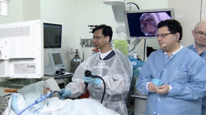
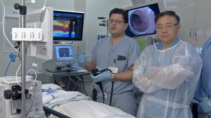
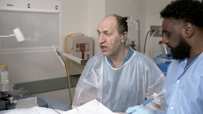
Live endoscopic procedures from the 11th Annual Long Island Live Endoscopy Course, Frontiers of Endoscopic Surgery. Part one of two, morning session.

Related Presenters

Stavros N. Stavropoulos, MD.

Stavros N. Stavropoulos, MD

Education Medical School: Columbia University School of Nursing Residency: Columbia University Medical Center Fellowship: Columbia University Medical Center Board Certifications AB Internal Medicine (Gastroenterology)

View full profile

Ping-Hong Zhou, MD.

Ping-Hong Zhou, MD

Doctor of MedicineGraduate SupervisorDeputy Director of Endoscopy CenterAssociate Professor of General SurgeryYouth Member of Chinese Society of Digestive EndoscopyMember of Endoscopy Branch of Chinese Medical Doctor AssociationMember ...

View full profile

Haruhiro Inoue, MD.

Haruhiro Inoue, MD

MD Degree from Yamaguchi University, School of Medicine (1977-1983) Qualification In Japan;MD (Issued in May 1983)Japanese National Board of General Surgery (Issued in April 1987)Japanese National Board of Digestive Surgery (Issued in ...

View full profile

ADVERTISEMENT

Related Videos