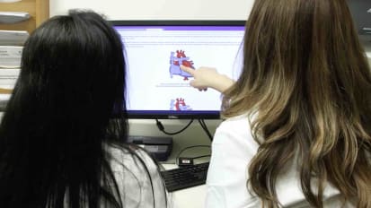
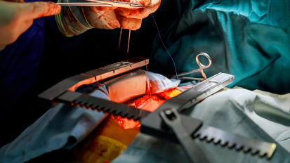
[MUSIC PLAYING]
VADIM FEDOROV: At the Ohio State University, we've developed a special program where we're collecting a live human heart. It's directly taken from the chest of the patient, diseased heart, and the patient will receive a new donor heart.
So we put the heart in the special ice solution, wash out from the blood. It's such the heart could be preserved for about 24 hours in this solution. Put these tubes inside of their main coronary arteries, which go in from aorta. As such, the heart could be perfused under 50, 60 millimeters of mercury, the pressure-- the same pressure which will beat in our heart.
Then heart warm up and start beating back. Again, it can beat in different rate because most of the hearts, they have different kinds of disease. After this heart is recovered, we put in all our electrodes, clinical electrodes, whilst we focus in our optical apparatus, different cameras. Then we can inject special fluorescent dye which can sense electricity.
After we inject this dye, and they put in red light, we excite this fluorescent probe. And we can see how electrical wave propagates across the heart, where is the origin of our electrical heartbeat, how it's propagated, and where is the problem. So we can see these arrhythmias from the optical cameras.
Actually, simultaneously, we can see 60,000 recordings across the whole heart and visualize whole electrical activity in our human heart in 3-D. And after that, we see where the source of this arrhythmia. We put in clinical catheter exactly on this spot. make this local ablation. We destroy only 3 by 3 millimeters, but it has to be, also, in the depths, too-- transmural ablation. And if we put this on the right spot, we completely eliminate this small [INAUDIBLE] re-entry micro re-entry activity.
However, our discovery, specific for the human heart, we call this micro-re-entry, micro-anatomic re-entry, Because we found they always have special connective tissue, fibrotic lesions. And the first activity develops around this connective tissue.
But we can see in 3-D, and we can see it's kind of a loop, provides very fast source of electrical activity in our heart. If we define where is this loop, we can put electrodes right there, ablate it, destroy it, patient will be free from arrhythmia.
From 50% to 20% patients only could be treated successfully long-term from these arrhythmias. So we're looking forward to significantly increase this percentage based on that mechanism which we uncovered in the human heart. We should be studying these high-resolution optical imaging, and this imaging can significantly improve patient-specific atrial fibrillation treatment.