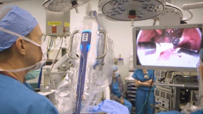
Adam Lustig, M.D., Interventional Radiologist at Medical Center Radiologists, discusses a case presentation of a prostate artery embolization on a 61 year old male who had an enlarged prostate and wanted a minimally invasive option.
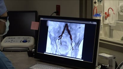
Hi, I'm Doctor Adam Lustig. I'm an interventional radiologist. I work for Medical Center radiologists or MCR, and primarily work at Centera Norfolk General and Centera Lee. Perform a variety of minimally invasive procedures on a variety of diseases. So I wanna talk to you today about. A case presentation of a prostate artery embolization I did a few months ago. This is a 61-year-old gentleman who has an enlarged prostate and lower urinary tract symptoms from his enlarged prostate. He wanted a non-surgical option, a minimally invasive option that didn't have quite the Level of potential complications that can come with surgery. So he came to me. He saw me in clinic at our Norfolk General office, and we talked to him about the potential options in the procedure, and he had an IPSS score, which is an International prostate symptom. Scale or the AUA score in the twenties. His was 24, which is pretty standard. The max is 35. He had pretty severe BPH and lower urinary tract symptoms, so I discussed with him the procedure, and I'd like to go over it with you today. So here this is the beginning of the procedure, getting access into the arterial system through the radial artery ultrasound guidance, and once we get flash, we advance the wire through the needle and get access into the radial artery. Over the wire we will place a sheath which is coming in right here and it's got a side port where we can get continuous saline being flushed through the sheath so it doesn't form a clot at the tip of the sheath. And now we have a way to advance and exchange catheters and wires safely into the arterial system. So through the radial artery advancing a flush catheter so we can take a good flush picture of the pelvic arteries in the aorta and advancing this using X-ray guidance in real time to see the catheter going down the aorta into the pelvis. And now we're doing a cone beam CT, which is a contrast injection through the catheter in the pelvic aorta while doing a CT scan on the table. This is a fairly new machine that we have at Norfolk General, and this is the result. We get a CTA of the pelvis. You can see the arteries have contrast in it, and I'm drawing a sort of border around the prostate which will help the computer determine which arteries are going and supplying the prostate. You can see me pointing to the left prostate artery there. And once we once we have a nice picture, I can cut out all the extraneous anatomy that I don't need like the pelvic bones and some other arteries and vessels that just aren't important for this case. After I cut that out, we get a nice vessel map, and you can see in the red and yellow lines are what the computer thinks are the prostate arteries, and I agree with the computer in this case. So using this in real time, we can overlay that on the real-time fluoroscopy which can Help guide the catheters and wires during the procedure, and what that does is reduce radiation, it reduces contrast usage, and reduces the amount of times I need to do a wire or catheter exchange to inject contrast, and this will rotate in real time if I move the gantry left or right around the patient, that 3D map will actually shift in real time. There you can see on the screen it's overlaid on the right side in sort of an oblique projection. So once we get to the prostate artery, we inject these particles that are 300 to 500 microns in size, and here I'm in the left prostate artery and injecting little particles that are going to actually embolize the prostate artery, and that's the actual crux of the procedure is injecting those tiny particles into the prostate artery to embolize the prostate tissue and ultimately shrink the prostate to relieve the patient's lower urinary tract symptoms. And so here I have a manifold of the big syringe on top in blue, and we load the smaller syringe in red with more particles, and that will be injected into the artery. Once we are done with one side, we go to the other side and repeat the same process. Those particles will travel pretty distally, which is what we want. We also want to occlude the main trunk of the prostate artery so that we can reduce recantalization of the artery in the future. And what I like to do is inject a little bit of gel foam, which is what is being mixed here, and the gel foam just has larger particles and there's a higher volume that you can inject into the main trunk of the prostate artery. And so that will embolize completely both prostate arteries and decrease significantly the blood flow to the prostate gland itself, which will over time allow the prostate to shrink. Compared to other procedures like TURP or prostatectomy, this will take several months for the prostate to shrink, and the patient may not and will not actually get immediate relief of their lower urinary tract symptoms because it will just take a few months for the prostate to shrink. But at the 6 month time point we have data that shows there's no statistical difference between the amount of improvement in lower urinary tract symptoms that a patient experiences between a TURP and a PAE or prostate artery embolization. So it's really just that time frame of waiting until the prostate shrinks and relieves that obstruction. Once we're done embolizing both sides, we have to pull out the sheath from the radial artery, and the way we close up the hole in the artery is by using a TR band, which is a wrist band that has a little pocket of air that we can inflate using a syringe with various amounts of air. And we deflate that to the point where there's still flow in the artery, so you can kind of see the pocket of air beating as the pulse is going through the artery, but we don't want it to be fully occlusive and we obviously don't want it to be leaking, so this is a standardized way to close a radial access. And that is the end of the procedure. So the patient will now stay for a few hours and go home the same day, and we'll see him back in clinic in a month. So this patient went home the same day as they typically do, and he followed up in my clinic in 1 month and then 3 months later, and his IPSS score has dropped both times, which is pretty typical of what we expect. He started out at a 24 and now he's around 10, and that's a pretty significant reduction, pretty typical for what you expect for a prostate artery embolization. His quality of life is much improved, and he's very happy. He had no complications, and this is sort of a pretty typical experience for a patient going through this procedure.
Related Presenters